
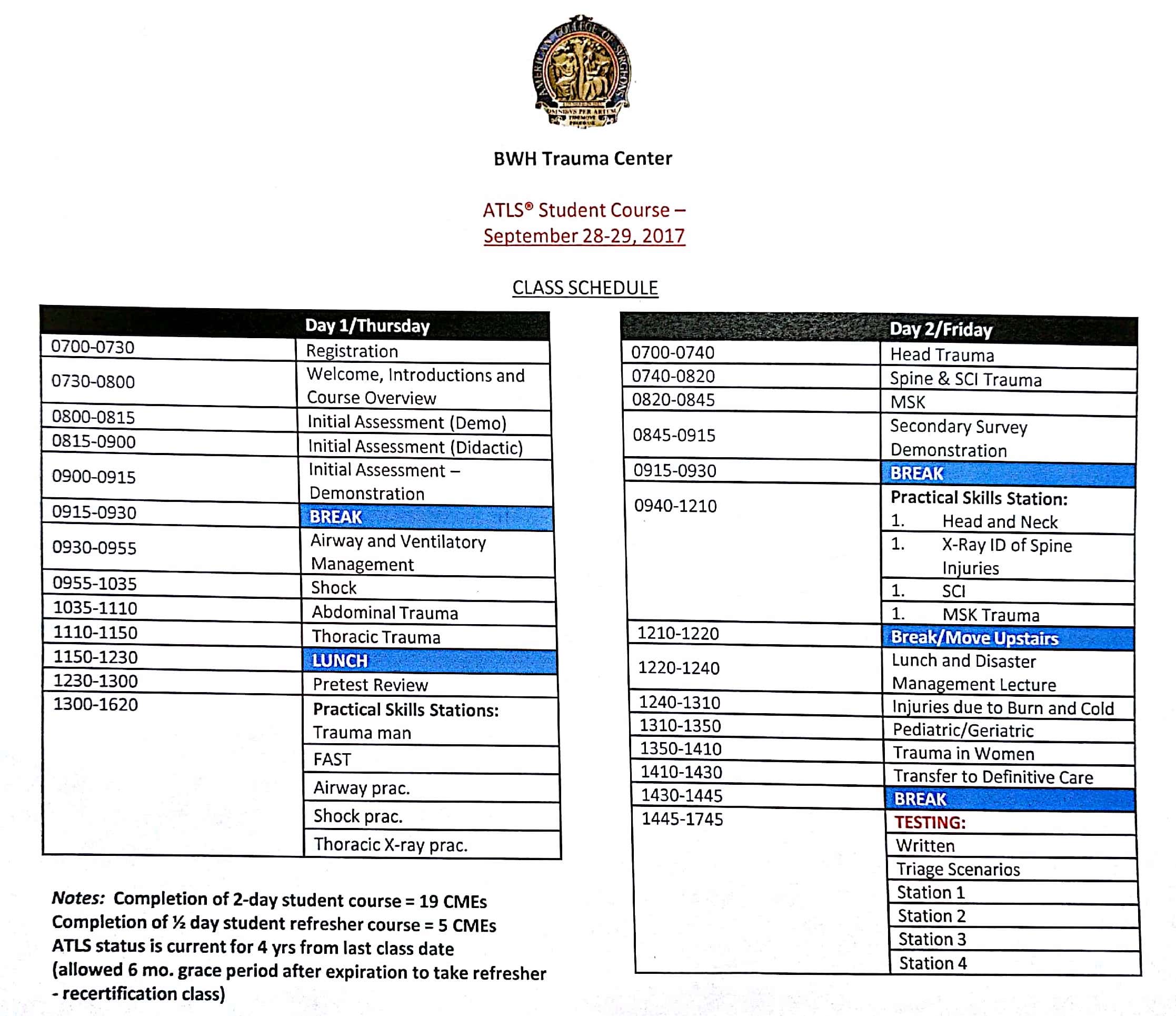
As of yesterday, I’m officially certified in Advanced Trauma Life Support (ATLS) for the next four years! This two day course was required and paid for by my critical care fellowship, so I wanted to summarize some of the pearls I learned.
In 1976, Dr. James Styner, an orthopedic surgeon, was piloting an aircraft with his wife and four children when it crashed in Nebraska instantly killing his wife and rendering three of his children unconscious. He and his eldest son transported the remaining children to a local hospital where Dr. Styner noted the lack of training in managing trauma patients.
“When I can provide better care in the field with limited resources than my children and I received at the primary facility, there is something wrong with the system and the system has to be changed.”
He became instrumental in developing ATLS which now serves as a common language in managing trauma and always starts with a primary survey remembered by A-B-C-D-E. This treats the greatest threat first, emphasizes the importance of time, and does NOT seek to establish a definitive diagnosis in the acute setting. Caretakers involved in ATLS should always practice standard precautions consisting of a cap, gown, gloves, mask, shoe covers, and face/eye shield.
- A: airway with cervical spine (C-spine) precautions
- occult airway injury, progressive loss of airway, equipment failure, inability to intubate
- B: breathing and ventilation
- respiratory rate, chest movement, air entry, hemoglobin oxygen saturation
- be wary of positive pressure ventilation converting an iatrogenic pneumothorax into a tension pneumothorax
- C: circulation and hemorrhage source control
- assess for organ perfusion – level of consciousness, skin color, temperature, pulses
- control hemorrhage – restore volume, apply pressure/tourniquets, reassess for appropriate response, perform a focused assessment by sonography in trauma (FAST)
- D: disability and neurological exam
- obtain a baseline neuro exam, GBS, and pupillary response
- important to perform a quick neuro exam prior to intubation as sedation/paralytics will confound the exam
- E: exposing the patient, ensuring a safe environment
- completely undress patient, find missed injuries
- prevent hypothermia
- make sure the patient’s environment is safe
The primary survey is repeated frequently to identify any deterioration in the patient’s status indicating the need for additional intervention. As adjuncts to the primary survey, we should consider adjuncts EKG, blood gases, labs, urinary/gastric catheters, and x-rays of the chest and pelvis. Next, perform the secondary survey, a complete history and physical (H&P) with head-to-toe physical exam.
- History (AMPLE) – allergies, medications, past illnesses/pregnancy, last meal, events/environment of trauma
- Head: scalp palpation (potential bleeding), comprehensive ear/eye exam, periorbital edema, occluded auditory canal
- Maxillofacial: bony crepitus, deformity, malocclusion, potential airway obstruction, cribriform plate fracture
- Neck (soft tissues): blunt vs penetrating trauma, airway obstruction, hoarseness, crepitus, hematoma, stridor, bruit
- Chest: inspect, auscultate, palpate, x-ray, E-FAST exam
- Abdomen: inspect, auscultate, palpate, E-FAST, hollow viscous injury, retroperitoneal injuries missed with E-FAST
- Pelvis: leg length unequal, instability, pelvic x-ray as needed, potential for large blood loss and damage to genitourinary system, place pelvic binder and only examine once if concerned about pelvic instability
- Perineum/rectum: contusions, hematoma, sphincter tone, high-riding prostate, urethral/rectal blood
- Extremities: contusion, deformity, pain, perfusion, neurovascular status, x-rays as needed
- Neurologic: GCS, pupil size/reactivity, lateralizing signs, early neurological consultation and prevent secondary injury
If the provider feels that their facility is not equipped to handle the clinical situation at hand, arrangements must be made ASAP for transfer to a hospital with the necessary accommodations. Transfer should occur after stabilizing measures (securing the airway, hemorrhage control) but not be delayed by adjuncts like imaging.


