Ultrasound is not only a great bedside diagnostic modality, but it’s routinely used to guide procedures like line placement, peripheral nerve blocks, and thora/paracentesis. It relies on pulses of high frequency sound waves reflecting off structures of varying acoustic properties to generate echos that are subsequently assembled into an image.
The piezoelectric effect is the cornerstone of traditional ultrasound. This is an electromechanical property of certain materials like quartz where an electrical current applied through the object generates vibrations resulting in pulsed sound waves. In turn, echos reflected back on the crystal generate changes in electrical resistance and current. In short, the conversion of electrical energy (a current applied over a voltage) to mechanical energy (sound waves) is the key!
Now let’s talk about the sound waves themselves. As with any wave, its frequency x wavelength = constant velocity through a medium. Because we’re shooting ultrasound through a body cavity which contains blood, muscle, fat, bone, etc., the speed will vary through these different tissues. Every time the sound waves pass through a different medium, the beam is rarefracted and some of the waves can change direction/velocity. The ultrasound can only create an image with sound waves that are reflected back to the probe’s transducer. A computer analyzes the time delay between when the pulse was sent, when the echo was received, as well as properties of the sound itself (amplitude, pitch, etc) to calculate velocities and create images.
Typically, medical ultrasound operates at 2 – 27 MHz. That’s 2 to 20 MILLION oscillations per second. A high frequency ultrasound transducer (vascular linear probes), the tissue penetration decreases but image resolution improves. Likewise, a low frequency probe (large curvilinear probe) will have increased tissue penetration but poor image resolution. These properties are due to attenuation of the ultrasound signal as it traverses tissues of varying depth. Deeper tissues tend to have reflected waves with lower amplitudes. This is where the gain function comes in handy. By altering the time-gain compensation, we basically increase the signal of the reflected echos from deeper tissues to mitigate artifacts.
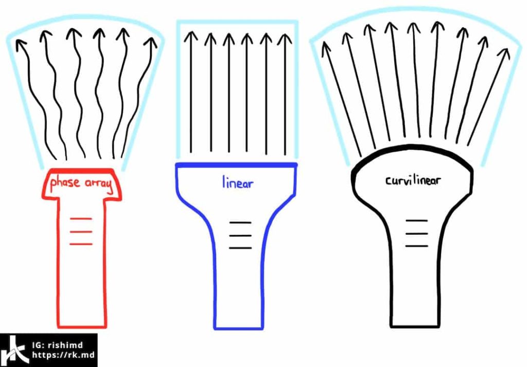
Here are three different types of ultrasound transducers. The flat-faced linear probe has crystals which are placed in parallel producing a rectangular field of view. These are traditionally the probes we use for vascular access, most peripheral nerve blocks, assessing superficial soft tissues, etc. The curvilinear probe projects sound waves widely in a curved fashion at low frequency making it ideal for intraabdominal imaging. Phase array probes also project widely but have many transducers which pulse waves independently with a certain time delay between each one. This results in constructive interference of each transducer’s sound waves at some new wavefront which is angled relative to the probe with reflections that are stitched together into an image.
Drop me a comment below with any questions! 🙂


Hello and thanks for your explanation. I have a question for you.
What characteristics do you think a good probe should have for examining nerves and muscles?
Thanks in advance
Depends on a lot of factors. If your structure of interest is superficial, higher frequency (usually linear probe) is the way to go. Deeper structures (subgluteal sciatic nerve) probably need lower frequency, curvilinear probes. There’s a lot of overlap, and ultimately one has to consider user preference too.
Thank you soo much! You have no idea how much this helped me (and others)!
My sincere privilege, Nacole! 🙂
Hi Dr. Rishi, first of all congratulations for ur activities on ig and on this blog. I would ask what’s the use of the phase array probe?
Thanks.
Hey Savi! Thanks for the kind words! Phase array probes are incredibly useful for assessing deeper structures through small acoustic windows (ie, the small window between ribs when looking at the heart or lungs).