In this portable x-ray of a patient with COVID-19 pneumonia, one can clearly see the right and left mainstem bronchi as well as more distal segments (arrows indicate several “air bronchograms”). This occurs because the air-filled, patent bronchi don’t attenuate x-ray radiation and appear dark compared to the surrounding airspace diseased from fibrosis, consolidation, infection, edema, hemorrhage, etc. The patent airway anatomy (black) is accentuated on the opacified (white) airspace disease suggesting that something other than air is filling the alveoli.
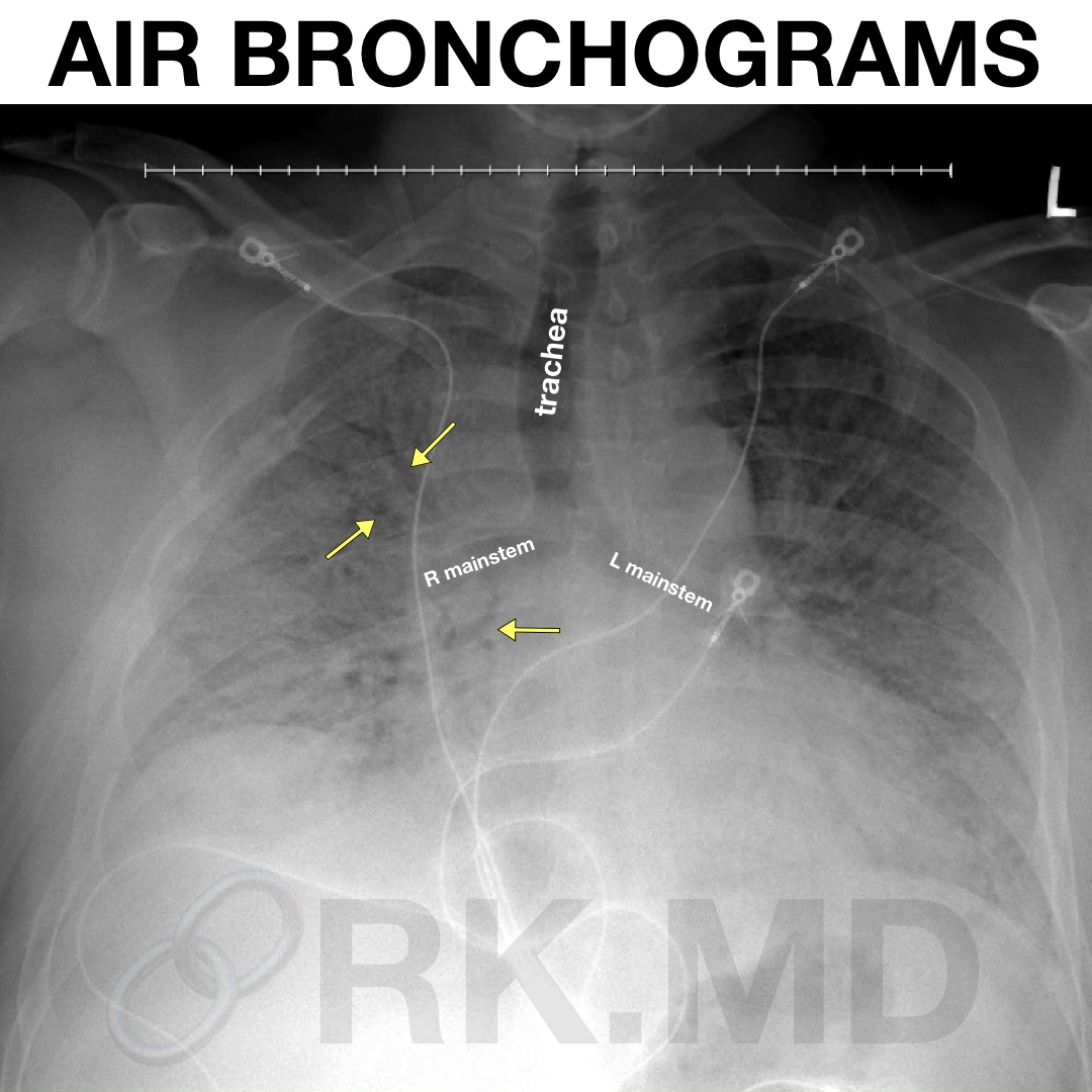
On a portable x-ray, it’s often difficult to differentiate atelectasis (parts of the lung which have collapsed) from consolidation (lobar pneumonias, etc.) Airspace consolidation usually has air bronchograms whereas atelectasis usually does not, but as with anything, clinical correlation and other techniques like bedside lung ultrasonography are important considerations!
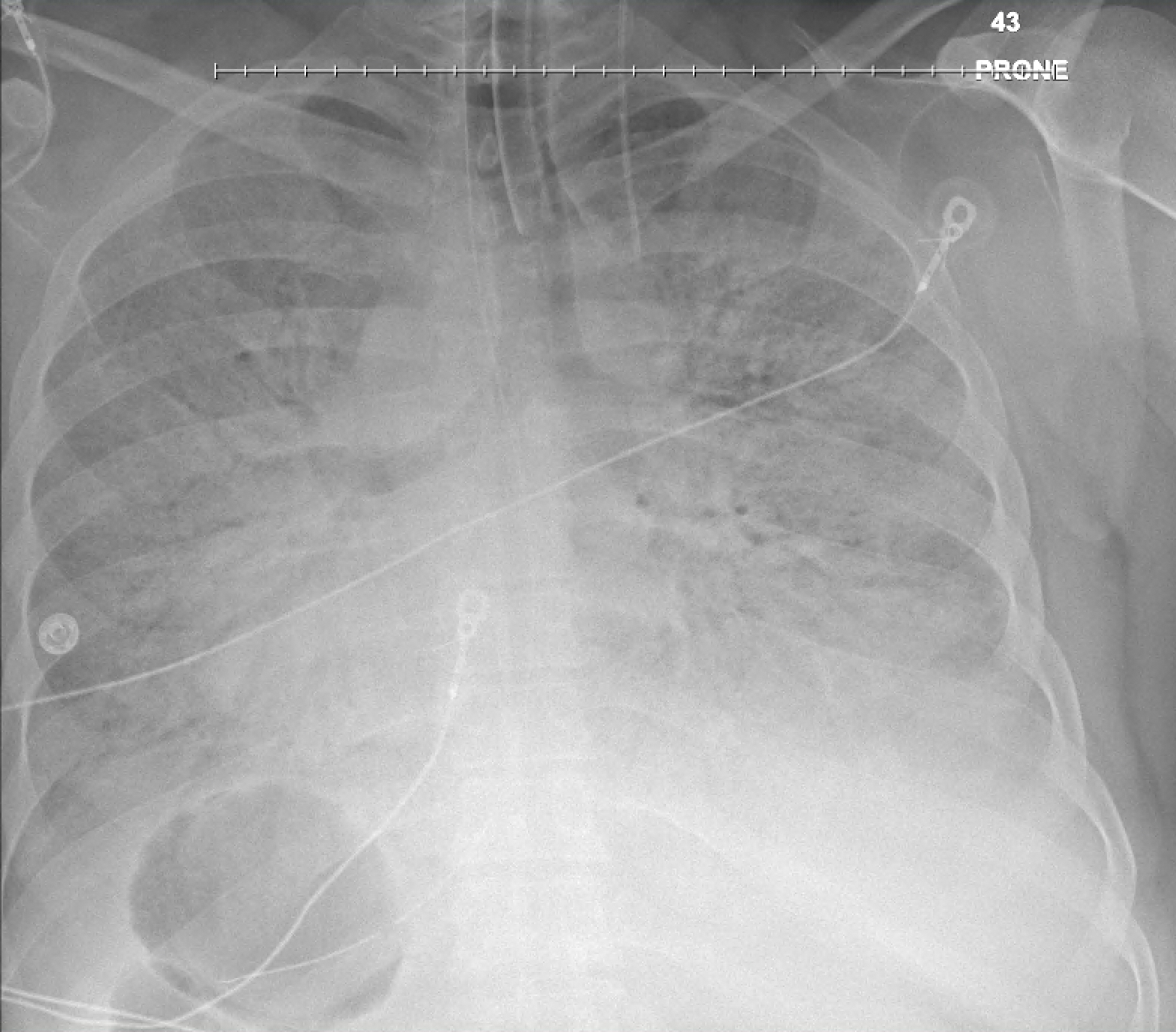
Here’s another example of a chest x-ray in the prone position (patient on their abdomen) in severe COVID-19 pneumonia. Notice all the air bronchograms everywhere!