
For patients with difficult arterial access (namely calcified/narrowed femoral and subclavian arteries), a transcaval approach offers another alternative. In this technique, interventional cardiologists guide a catheter through the femoral vein into the abdominal inferior vena cava (IVC). Using prior CT imaging and intraprocedural fluoroscopy, they electrify a venous guidewire from the IVC into the aorta where it’s essentially “caught” by a snare and advanced retrograde into the aortic root to perform the TAVR in the conventional manner.
Yes, there’s a hole between the IVC and abdominal aorta in the retroperitoneal space bridged by the TAVR delivery catheter. After the procedure is finished, the aorta is typically closed with an Amplatzer device while the hole in the inferior vena cava is left open. The thought is that the pressure within the retroperitoneal space (~20 mmHg) exceeds that of the venous pressure in the IVC (~10 mmHg). This pressure gradient suggests that blood should remain intravascular.
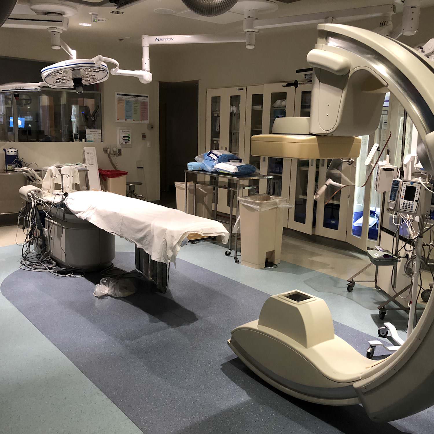
As an anesthesiologist, I still attempt my routine TAVR anesthetic; however, in addition to the considerations associated with TAVR (heart block, hypotension, loss of pacing, aortic root disruption, coronary artery occlusion, peripheral vascular injury, etc.), I must always consider the potential for retroperitoneal hemorrhage. You can hide a lot of volume in this space, and since our patients are supine for the procedure, physical signs such as bruising may not be apparent. Any form of hemodynamic lability refractory to conventional interventions should entertain the need for a stat CT or venogram to look for extravasation.


