• Subaortic Membrane

    Subaortic Membrane

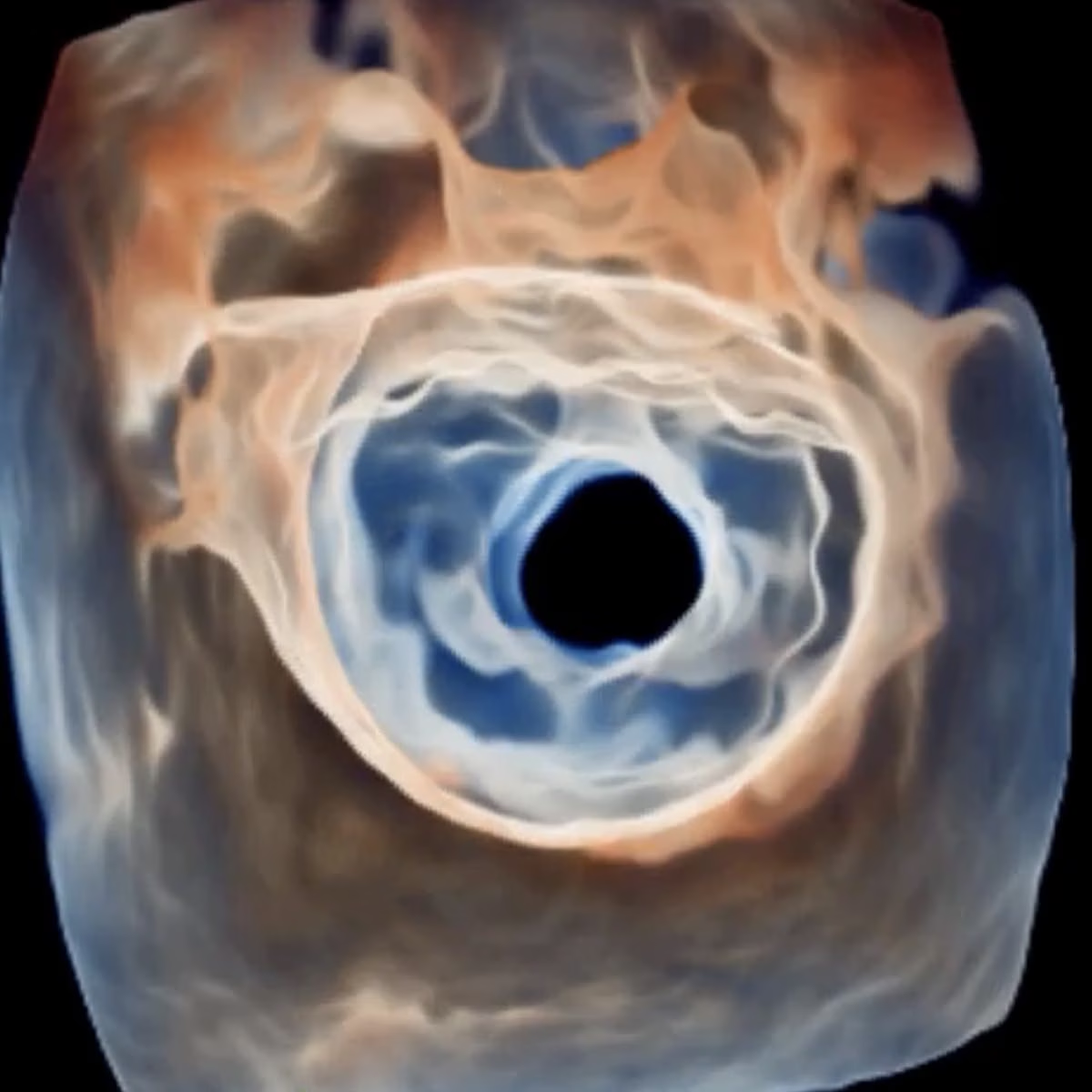
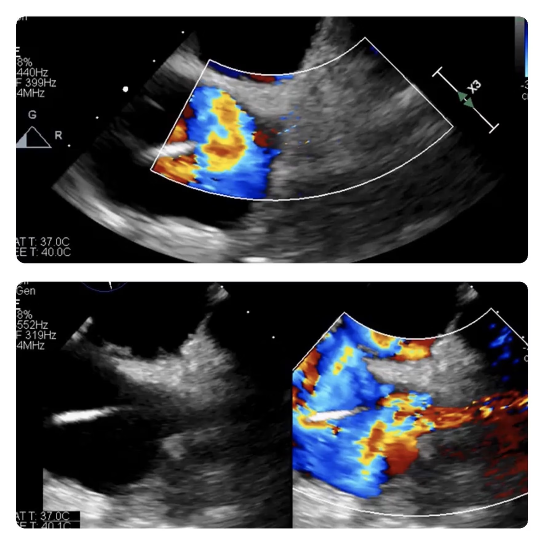
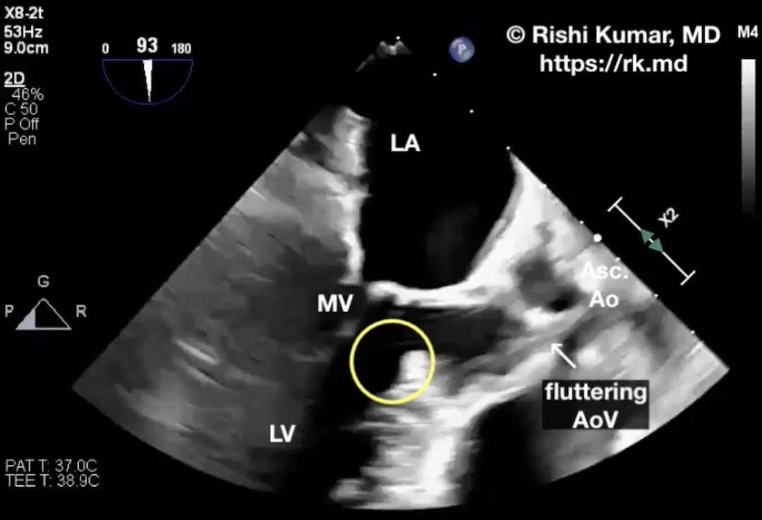
    Here’s a transesophageal echo (TEE) of a subaortic membrane (encircled in yellow) causing left ventricular outflow tract (LVOT) obstruction. Also note the fluttering of the aortic valve (AoV) leaflets during systole, turbulent flow in the LVOT, and chordal systolic anterior motion (SAM) of the mitral valve (MV). Surgeons will resect…
    Read More

  • Ocular Ultrasound For Increased Intracranial Pressure

    Ocular Ultrasound For Increased Intracranial Pressure

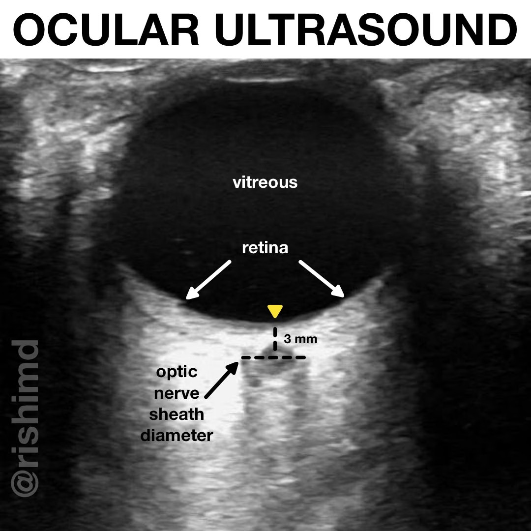
    As part of a quick point-of-care ultrasound (POCUS) examination, ocular ultrasound can help determine increased intracranial pressure (ICP), retinal detachment, lens dislocation, vitreous hemorrhage, etc. in conjunction with the patient’s history and physical exam.

    Read More

  • M-Mode Ultrasound Assessment Of Diaphragm Movement

    M-Mode Ultrasound Assessment Of Diaphragm Movement

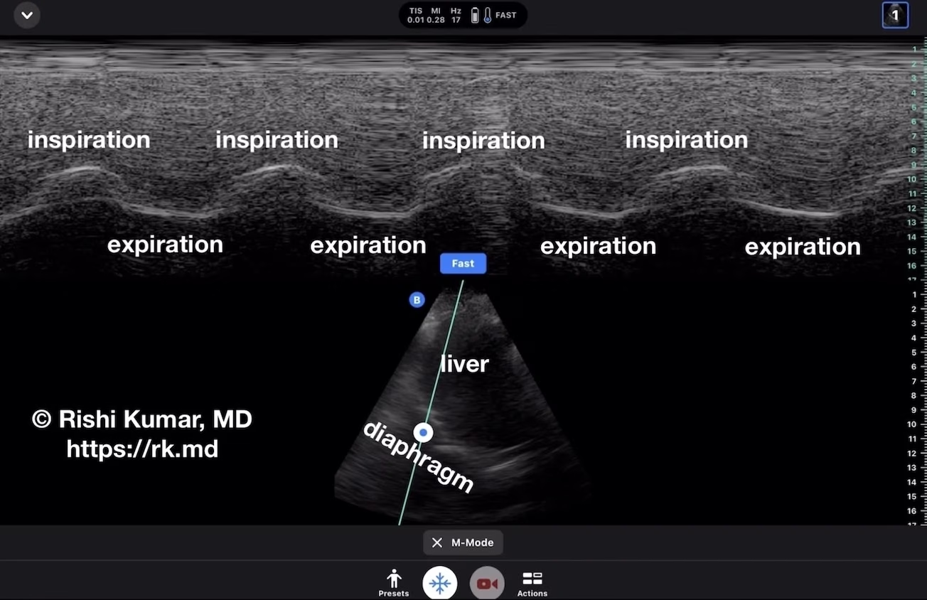
    Patients with suspected diaphragm paralysis (e.g., an elevated hemidiaphragm on a chest x-ray, a surgery that is high-risk for compromising the phrenic nerve, etc.) can be evaluated using motion mode (“M-mode”) ultrasonography at the bedside. M-mode tracks the motion of tissue along a linear path over time. In this video,…

    Read More

  • National Board of Echocardiography (NBE) Advanced PTEeXAM Case Log Template

    National Board of Echocardiography (NBE) Advanced PTEeXAM Case Log Template

    Here’s an Excel template for those logging transesophageal echocardiograms (TEE) on the supervised training pathway for the National Board of Echocardiography’s (NBE) advanced perioperative TEE diplomate status. This template is based on the sample provided by the NBE. Please leave me a comment with suggestions or if you found this…

    Read More

  • Tendyne Transcatheter Mitral Valve Replacement (TMVR)

    Tendyne Transcatheter Mitral Valve Replacement (TMVR)

    Abbott’s Tendyne transcatheter mitral valve replacement (TMVR) provides an alternative to MitraClip for patients with symptomatic, moderate-to-severe mitral regurgitation who are not suitable candidates for traditional, open surgical mitral valve replacement. The ongoing, multicenter, international SUMMIT Trial will assess outcomes over the next few years. After preoperative planning, the Tendyne…

    Read More

  • POCUS – IVC Versus Aorta

    POCUS – IVC Versus Aorta

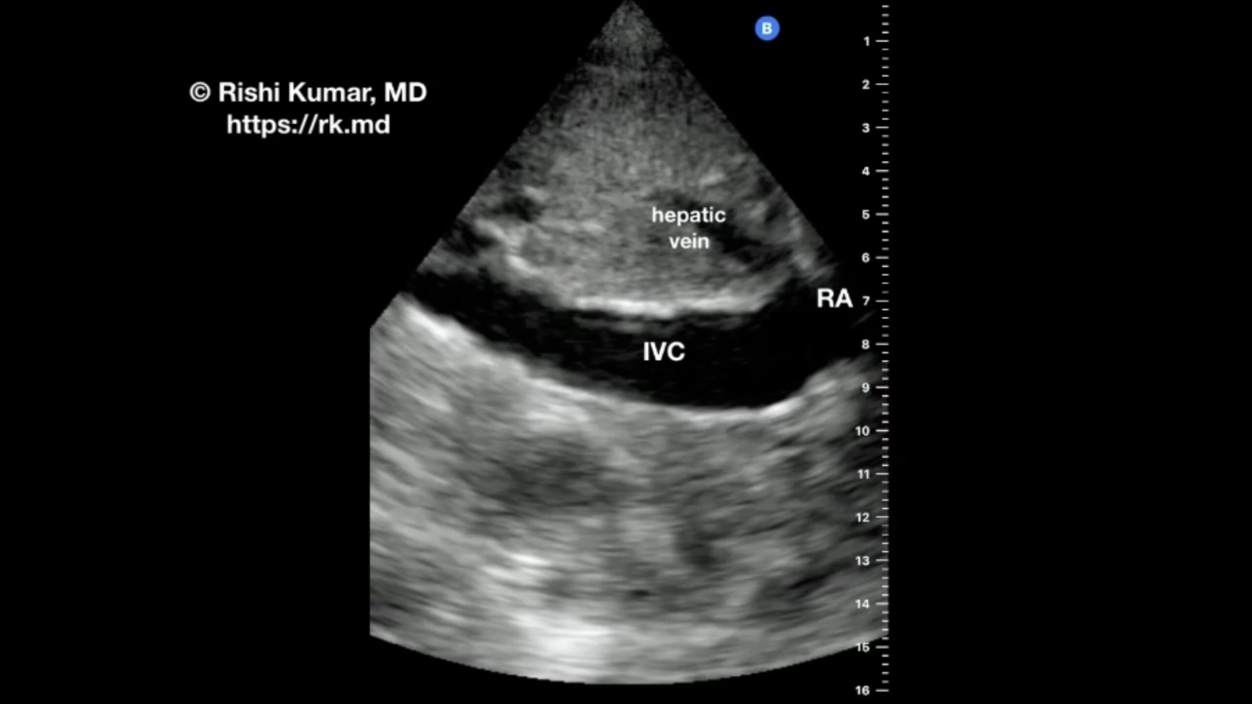
    The subcostal view of the inferior vena cava (IVC) is used to assess IVC diameter and respirophasic variation to gauge “volume status.” However, in my clinical practice, I find this technique fairly useless outside of the extremes. For example, an IVC diameter of 28 mm makes me less inclined to…

    Read More

  • Launch Of The Butterfly iQ+

    Launch Of The Butterfly iQ+

    As a cardiothoracic anesthesiologist and intensivist, I utilize ultrasound in a myriad of ways. Whether I’m placing a peripheral nerve block or performing arterial cannulation in the operating room or guiding a thoracentesis procedure or eFAST exam in the ICU, having ultrasonography at my disposal allows me to better care…

    Read More

  • Wall Motion Abnormalities (WMA)

    Wall Motion Abnormalities (WMA)

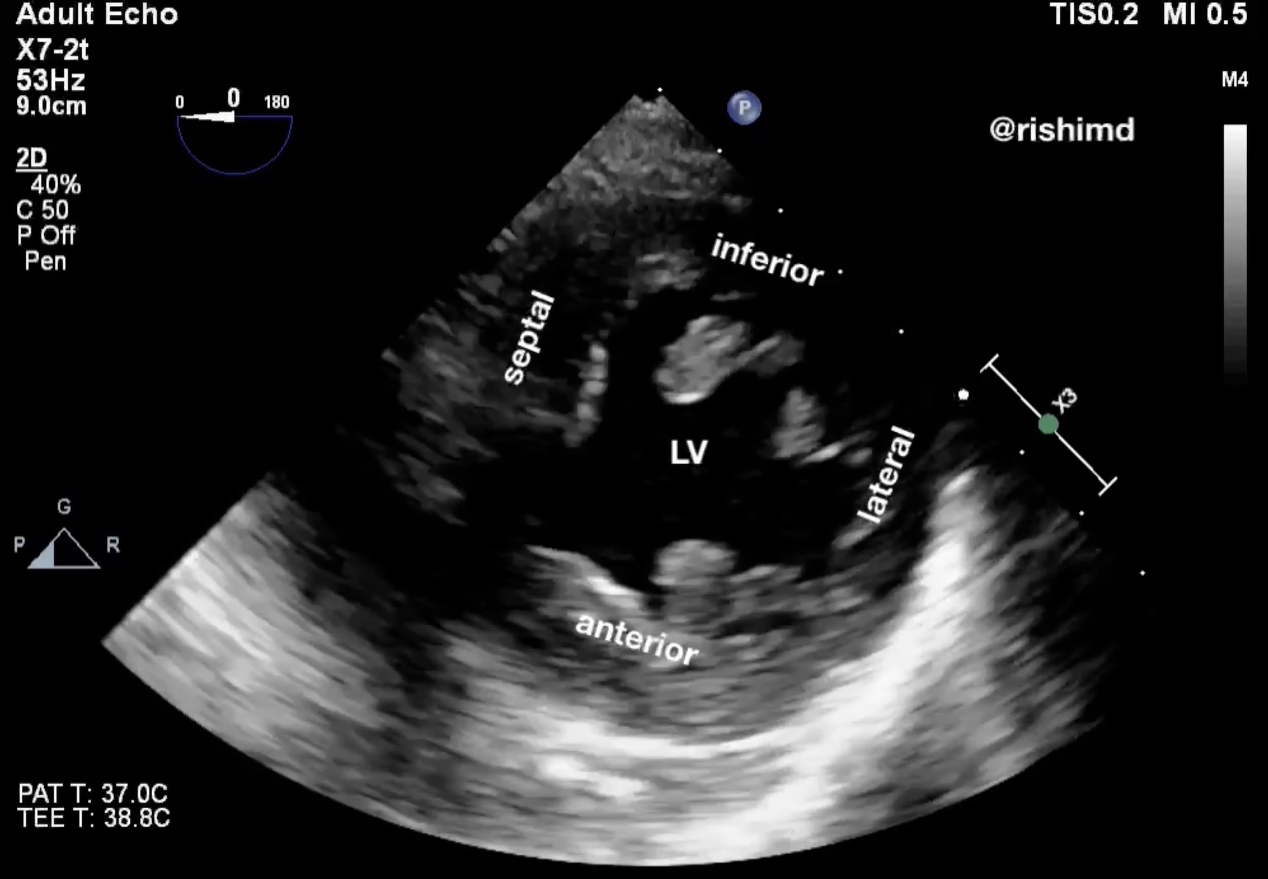
    Left ventricular (LV) wall motion abnormalities (WMA) are associated with myocardial ischemia and can be generalized or regional. In this transgastric mid-papillary short-axis view of the LV, one sees a clear difference in how the inferior and lateral walls are contracting compared to the septal and anterior walls. Decreased inward…

    Read More

  • Transposition Of The Great Arteries (TGA)

    Transposition Of The Great Arteries (TGA)

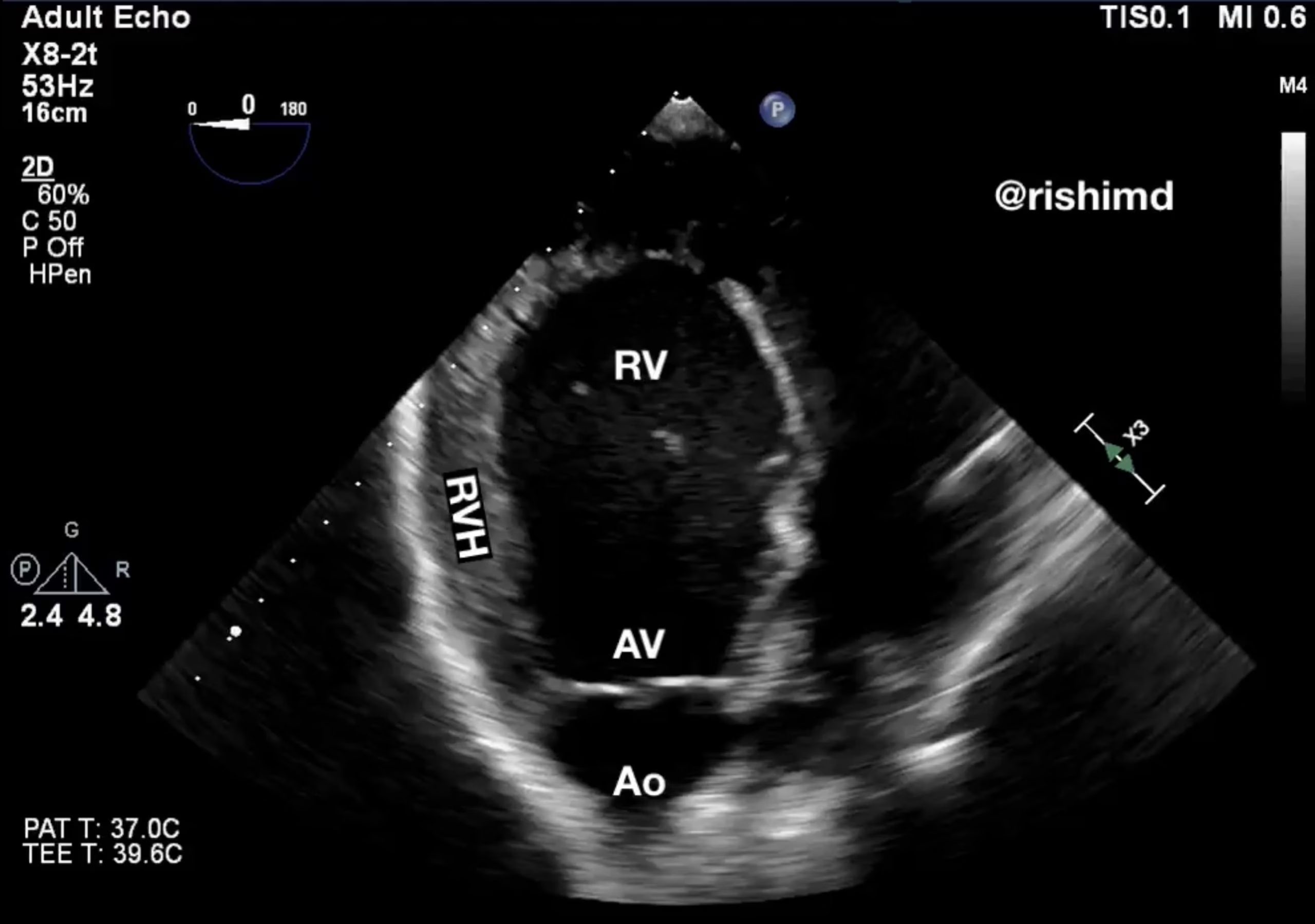
    Here’s a transesophageal echo (TEE) clip I’ve taken of an interesting congenital condition: transposition of the great arteries (TGA). Whereas the aorta (Ao) normally comes off the left ventricle (LV), in this case, it comes off a very hypertrophied right ventricle (RV). TGA comes in two variants. D-TGA has atrioventricular…

    Read More

  • Superior Vena Cava (SVC) Syndrome

    Superior Vena Cava (SVC) Syndrome

    Superior vena cava (SVC) syndrome is characterized by facial plethora, upper body venous engorgement, chest pain, soft tissue swelling, dyspnea, and even neurologic changes. Anything that impairs SVC drainage by intrinsic obstruction (thrombosis, anatomic narrowing) or extrinsic compression (mediastinal masses, non-small cell lung cancer, non-Hodgkin’s lymphoma, etc.) can result in…

    Read More