• Lung Point For Pneumothorax Using Ultrasound

    Lung Point For Pneumothorax Using Ultrasound

    With a pneumothorax (PTX), air separates the visceral and parietal pleura layers located on the lung’s outside surface and inner chest wall, respectively. The absence of pleural sliding on ultrasonography can suggest PTX, but when this is found adjacent to a lung point, it’s 100% specific for a PTX. The…
    Read More

  • D-Sign: Right Ventricular Strain On Chest CT And Echocardiography

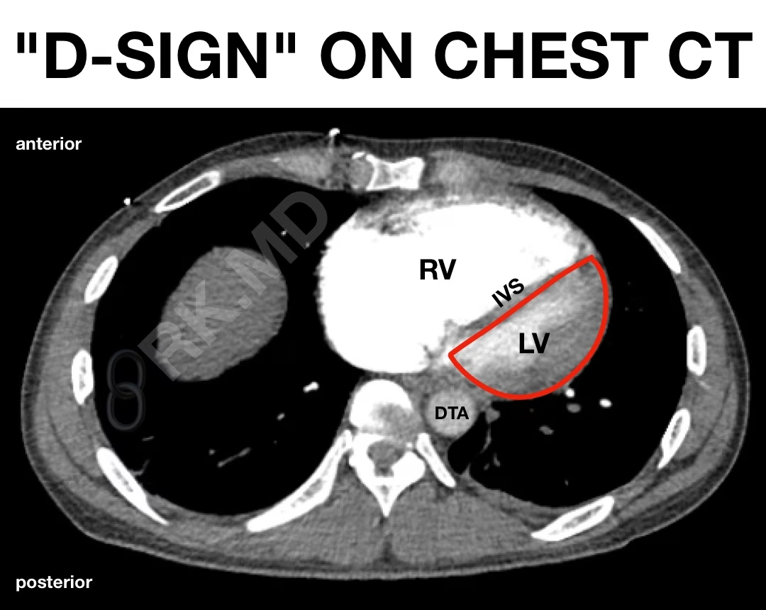
    D-Sign: Right Ventricular Strain On Chest CT And Echocardiography

    Typically at end-systole, the interventricular septum (IVS) bows rightward as the left ventricle’s pressure (LV) exceeds that of the right ventricle (RV). However, when the RV is strained (e.g., massive pulmonary embolism), the pressure overload can lead to flattening or even paradoxical bowing of the IVS. In a short-axis view…

    Read More

  • My Transesophageal Echocardiography (TEE) Sequence Of Views

    My Transesophageal Echocardiography (TEE) Sequence Of Views

    As a cardiothoracic anesthesiologist and intensivist, transesophageal echocardiography (TEE) is a powerful cardiac imaging modality I perform in the OR during cardiac surgery and in the cardiovascular ICU when point-of-care ultrasound is not feasible or helpful due to poor image quality. Before anything, I take a quick shot of the…

    Read More

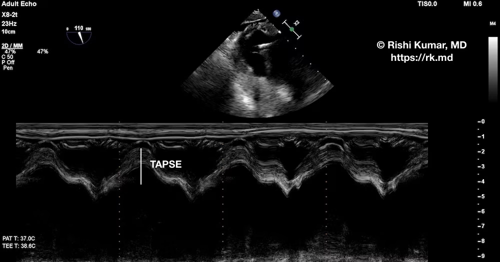
  • TAPSE With TEE

    TAPSE With TEE

    Tricuspid annular plane systolic excursion (TAPSE) is a quantitative echocardiography technique to assess right ventricular (RV) systolic function. As an intensivist, I obtain an apical four-chamber view using transthoracic echocardiography, activate motion (“M”) mode through the lateral tricuspid valve (TV) annulus, and measure the peak-to-trough distance. A value > 18…

    Read More

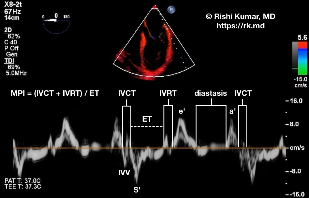
  • Myocardial Performance (Tei) Index With Doppler Tissue Imaging (DTI)

    Myocardial Performance (Tei) Index With Doppler Tissue Imaging (DTI)

    As part of a comprehensive left ventricular (LV) diastolic assessment, doppler tissue imaging (DTI) of the medial and lateral aspects of the mitral valve (MV) annulus should be performed. In the image below, I have labeled a DTI of the lateral aspect of the MV derived from a mid-esophageal 4-chamber…

    Read More

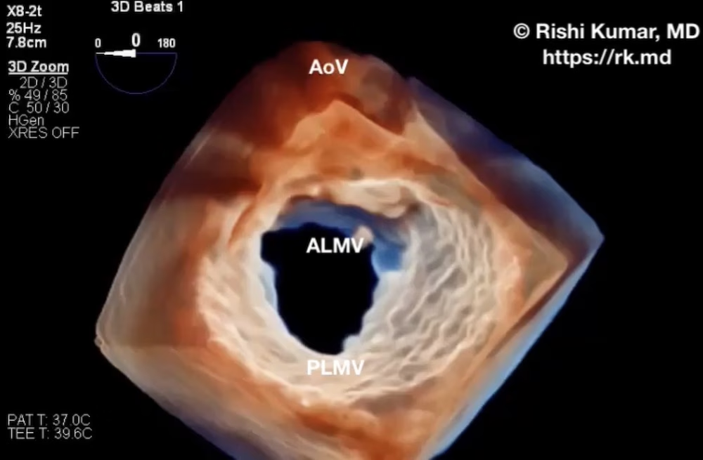
  • Mitral Valve Leaflet Perforation

    Mitral Valve Leaflet Perforation

    Mitral valve endocarditis can affect any part(s) of the mitral valve apparatus – annulus, leaflets, and even chordae tendinae – resulting in acquired mitral valve regurgitation (MR) through leaflet perforation, rupture of chordae, and other damage to the leaflets. 3D transesophageal echocardiography (TEE) is incredibly powerful in diagnosing the mechanism…

    Read More

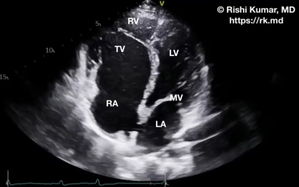
  • Ebstein Anomaly

    Ebstein Anomaly

    Ebstein anomaly is a rare (~0.005% of the general population) congenital heart condition characterized by the apical displacement of the septal tricuspid valve (TV) leaflet, a “sail-like,” long anterior TV leaflet, atrialization of the proximal right ventricle (RV), and a dilated TV annulus. These anatomic changes result in tricuspid regurgitation…

    Read More

  • National Board of Echocardiography (NBE) Critical Care Echocardiography (CCEeXAM) Case Log Template
  • Carcinoid Heart Disease Echocardiography

    Carcinoid Heart Disease Echocardiography

    Carcinoid syndrome is most commonly caused by an overproduction of hormones from metastatic midgut tumors and is characterized by diarrhea, flushing, hypotension, and increased airway reactivity. Roughly half of these cases also have carcinoid heart disease associated with fibrous deposition in right-heart structures. In particular, tricuspid and pulmonic valve leaflets…

    Read More

  • Left Atrial Myxoma

    Left Atrial Myxoma

    Myxomas are the most common primary, benign cardiac tumors occurring. They typically occur in middle-aged adults with a higher incidence in women. Most commonly, myxomas are located in the left atrium (LA). Large myxomas can cause obstruction of the mitral inflow leading to hemodynamic changes similar to mitral stenosis. Smaller…

    Read More