
The Couinaud classification system divides the liver into eight functionally independent segments, each with a center consisting of a portal vein, hepatic artery branches, and bile duct drainage. The periphery of each segment is where vascular outflow occurs via the hepatic veins. The numbering system begins with the caudate lobe (segment I, on the posterior of the liver and not seen in this image) and is carried out clockwise. Of note, segments VI and VII were drawn for the purpose of illustration. They will typically not be seen on an anterior view of the liver since they are posterior structures.
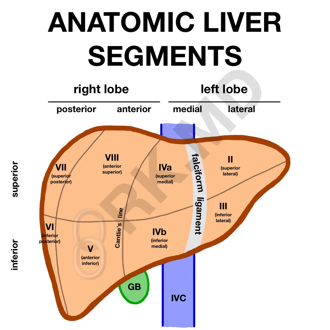
The right hepatic vein divides the right lobe into anterior (V and VIII) and posterior segments (VI and VII). The left hepatic vein divides the left lobe into lateral (II and III) and medial (IVa and IVb) segments. The middle hepatic vein divides the liver into right and left lobes. The portal vein divides the liver into superior and inferior segments.
The gallbladder (GB) separates segments V and IVb, and the falciform ligament divides the left lobe into medial and lateral segments (like the left hepatic vein).
Knowing the liver segments is helpful to localize pathology and discuss planes for surgical resection (e.g., extended right hepatectomy, left medial sectionectomy, etc.)
Drop me a comment below with questions! 🙂


Do the liver lobes have different functions?