
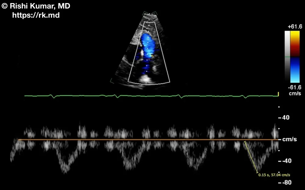
Pulmonary velocity acceleration time (PVAT), measured in milliseconds (ms), can be used to estimate pulmonary artery pressure (PAP) when a tricuspid regurgitation jet is difficult to locate or cannot be accurately interrogated with Doppler.
In this parasternal short-axis transthoracic echo view directed anteriorly and to the patient’s right, pulsed-wave Doppler is used to quantify the flow through the pulmonic valve. The time interval between the onset and peak flow velocity is the PVAT (0.15 seconds or 150 ms in this case).
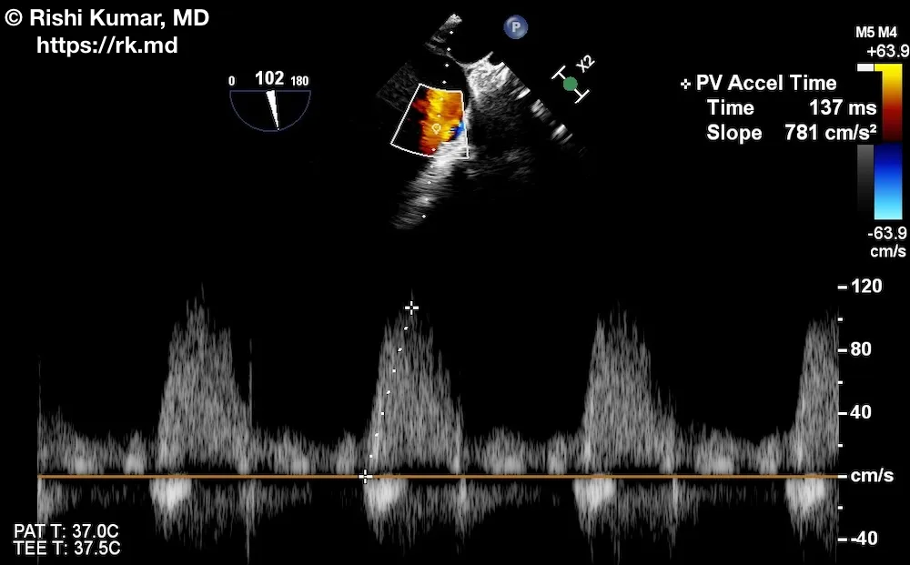
Here is a long-axis view of the pulmonic valve with transesophageal echo (flow directed to the probe, so above the baseline) with Doppler showing a PVAT of ~137 ms.
The severity of PAP elevation can be correlated to PVAT as follows: normal PAP (> 130 ms), borderline PAP (100 – 130 ms), mildly elevated PAP (80 – 100 ms), and severely elevated PAP (< 80 ms)
In situations of increased right-sided flow (left-to-right atrial shunting, severe pulmonic valve insufficiency, etc.), PVAT will underestimate the severity of PAP. In cases of severe pHTN, decreased cardiac output will result in lower velocities across the pulmonic valve too.


Thank you