• Concentric Left Ventricular Hypertrophy

    Concentric Left Ventricular Hypertrophy

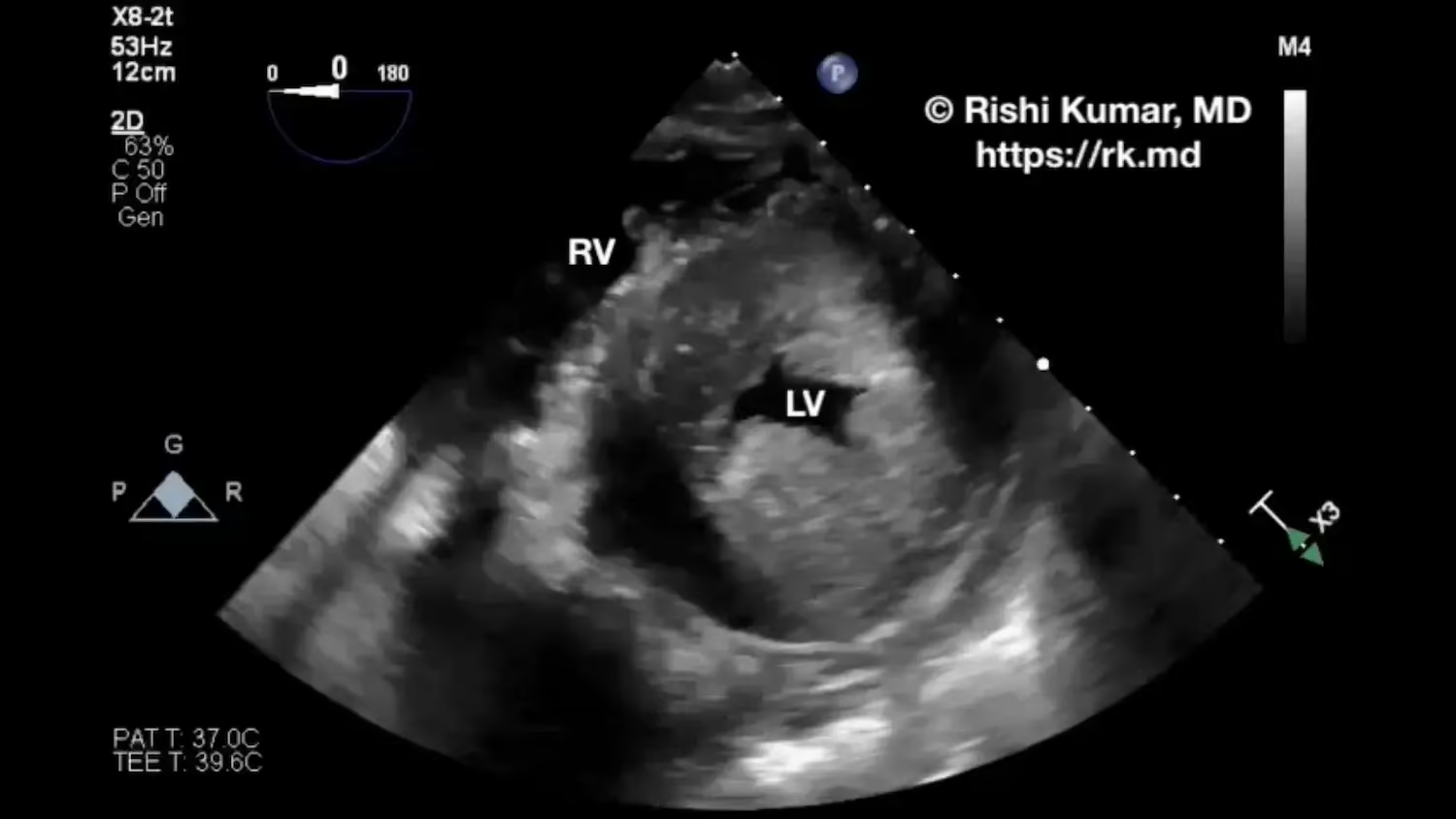
    Concentric left ventricular hypertrophy (LVH) is characterized by adaptive thickening of the myocardium in response to pressure overload from chronic hypertension or aortic valve stenosis. LVH can also be seen in genetic conditions like Fabry disease or hypertrophic cardiomyopathy with chaotic sarcomeric disarray at the cellular level. LVH leads to…

    Read More

  • Pulmonary Velocity Acceleration Time (PVAT)

    Pulmonary Velocity Acceleration Time (PVAT)

    Pulmonary velocity acceleration time (PVAT), measured in milliseconds (ms), can be used to estimate pulmonary artery pressure (PAP) when a tricuspid regurgitation jet is difficult to locate or cannot be accurately interrogated with Doppler. In this parasternal short-axis transthoracic echo view directed anteriorly and to the patient’s right, pulsed-wave Doppler…

    Read More

  • Cardiac Standstill POCUS

    Cardiac Standstill POCUS

    Point-of-care ultrasound (POCUS) can be extremely helpful during advanced cardiovascular life support (ACLS) with ongoing chest compressions and resuscitation. POCUS can help rule in or out certain etiologies (the H’s and T’s) for why a patient arrested. During the pulse check, I record the clearest subcostal 4 or 5 chamber…

    Read More

  • POCUS – Transhepatic IVC View

    POCUS – Transhepatic IVC View

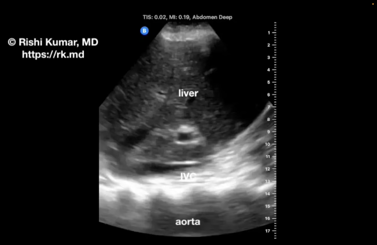
    The traditional way of assessing the inferior vena cava (IVC) with point-of-care ultrasound (POCUS) is longitudinally from a subcostal window; however, this can be challenging when that area is off limits (subcostal chest tubes, burn injuries, etc.) The transhepatic IVC view is obtained by placing a phased array probe (oriented…

    Read More

  • Carpentier Type III Mitral Regurgitation On TEE

    Carpentier Type III Mitral Regurgitation On TEE

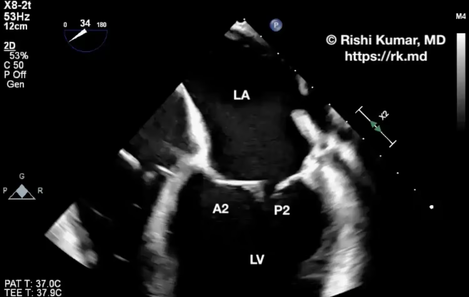
    Left ventricular (LV) dilation can stretch apart the mitral valve annulus causing the chordae tendinae to be pulled taut (particularly to the posterior leaflet). This restricts leaflet motion (Carpentier type III) over the cardiac cycle causing malcoaptation and associated mitral regurgitation (MR). Classically, with type III MR, the regurgitation jet…

    Read More

  • Surgical Septal Myectomy

    Surgical Septal Myectomy

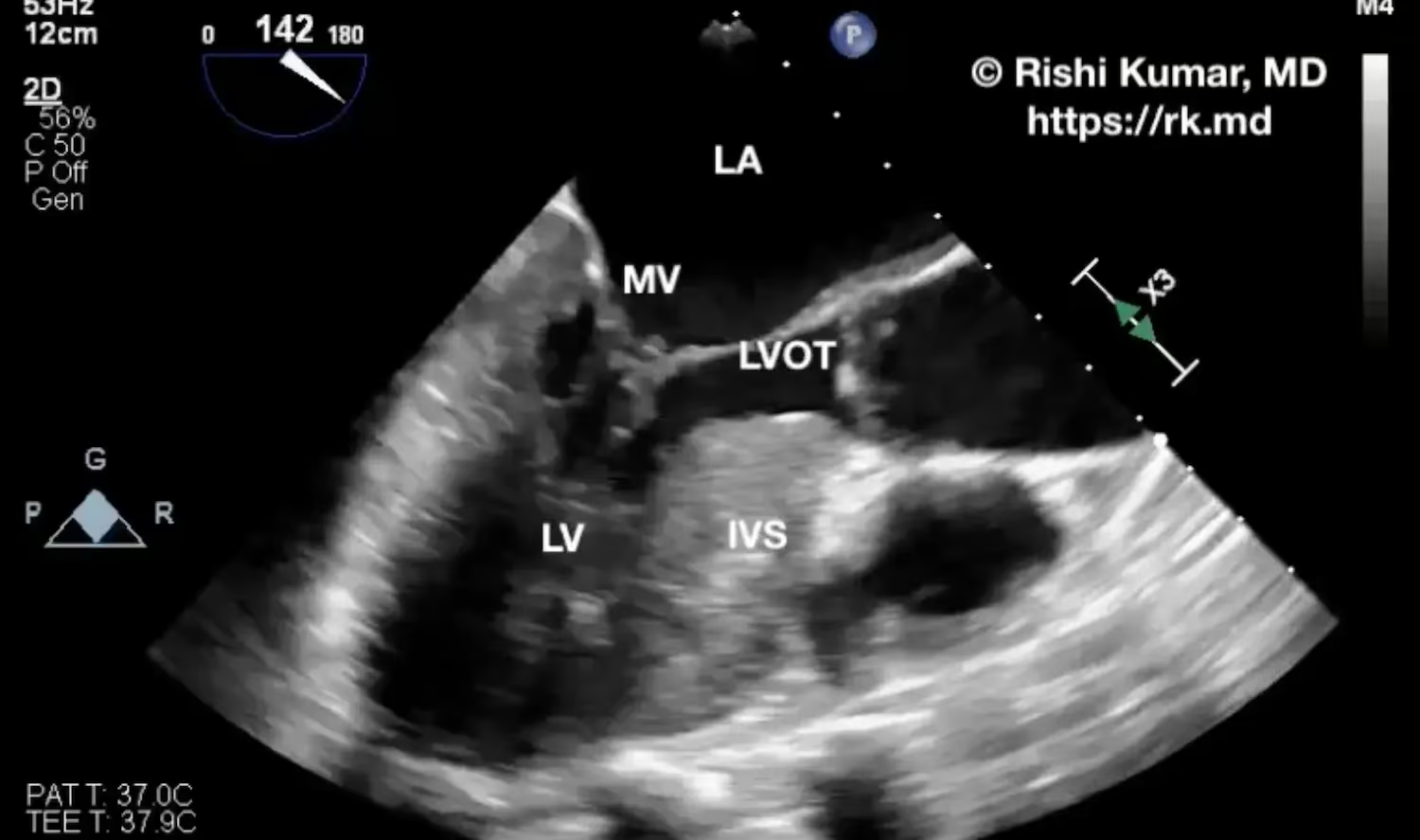
    Surgical septal myectomy is a procedure typically done for hypertrophic cardiomyopathy (HCM) where a portion of the hypertrophied interventricular septum (IVS) is resected. The basal portion of the IVS can lead to left ventricular outflow tract obstruction (LVOTO), limiting cardiac output and causing associated symptoms like dyspnea, lethargy, and organ…

    Read More

  • Air Bronchograms On Ultrasound

    Air Bronchograms On Ultrasound

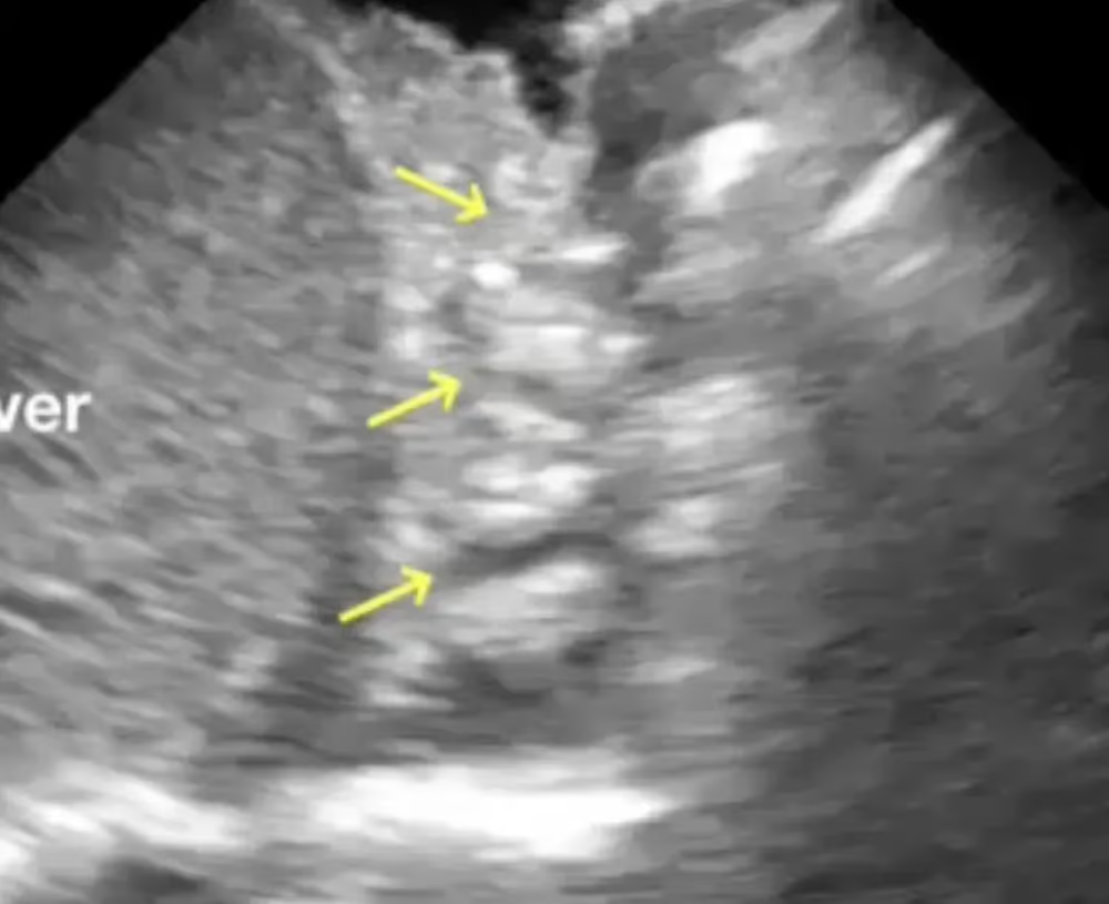
    In lung ultrasound, air bronchograms (indicated by yellow arrows in the video) occur when segments of the bronchial tree remain patent (filled with anechoic air) amid the surrounding consolidated, echogenic lung tissue. These adjacent alveoli could be collapsed as in atelectasis, filled with inflammatory exudates as in pneumonia, damaged in…

    Read More

  • 3 Point Compression Ultrasound (3PCUS) For Lower Extremity DVT

    3 Point Compression Ultrasound (3PCUS) For Lower Extremity DVT

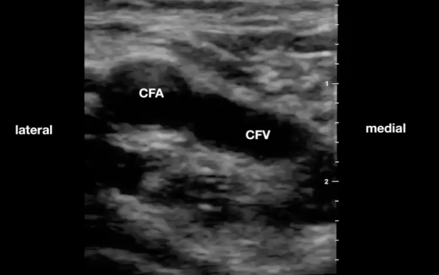
    As an intensivist, I’m always looking for ways to utilize ultrasonography to rule out/in pathologies on my differential. Rather than ordering lower extremity Doppler ultrasounds for every patient (a study that is costly from a time, financial, and human resource standpoint), I perform a three-point compression ultrasound (3PCUS) to rule…

    Read More

  • Doppler Shift – The Angle Matters!

    Doppler Shift – The Angle Matters!

    The Doppler shift equation describes the observed change in frequency or wavelength of a wave (such as sound, light, or radio waves) when the source of the wave and the observer are in relative motion. This phenomenon is known as the Doppler effect and is incredibly important to understand when…

    Read More

  • Tetralogy Of Fallot (TOF)

    Tetralogy Of Fallot (TOF)

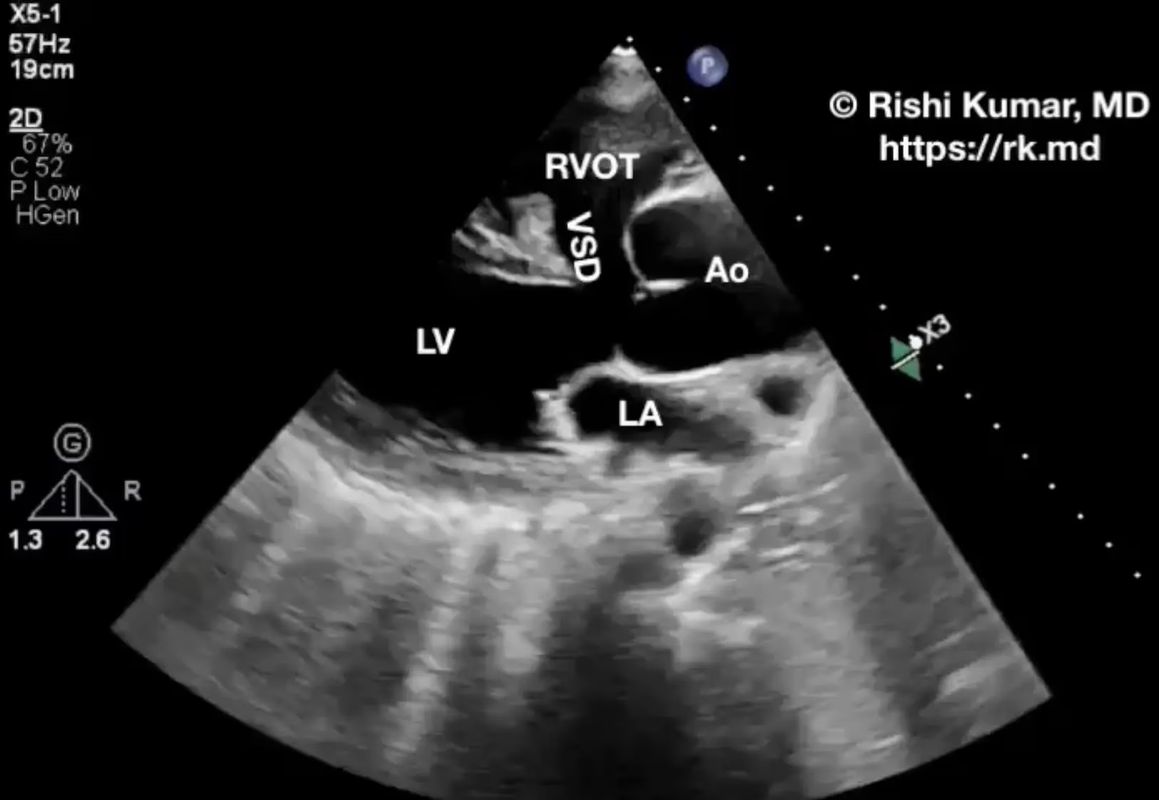
    Tetralogy Of Fallot (TOF), named for French physician Arthur Fallot, is the most common cyanotic congenital heart defect and comprised of four cardiovascular anomalies: RVOT obstruction reduces blood flow to the lungs resulting in chronic hypoxemia and right-to-left shunting through the VSD. It’s common for these patients to have hemoglobin…

    Read More