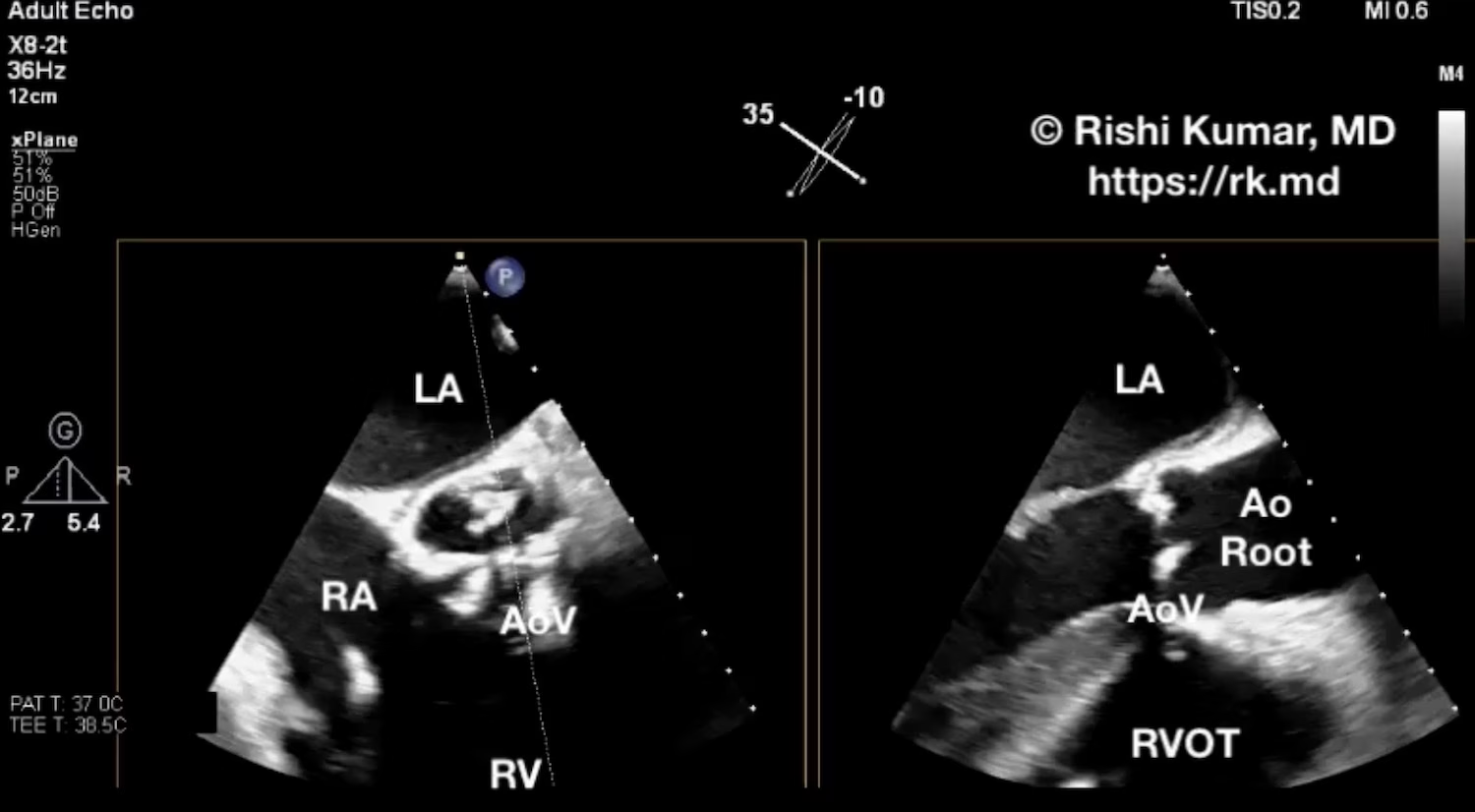
• Calculating Cardiac Output With Volume-Time Integral (VTI)

    Calculating Cardiac Output With Volume-Time Integral (VTI)

    Cardiac output (CO) is mathematically calculated as the product of heart rate and stroke volume. It’s related to preload, afterload, and contractility. One echocardiographic technique I routinely use to estimate a patient’s stroke volume is the volume-time integral (VTI). The VTI method is based on the Doppler effect which relates…
    Read More

  • Spinal Cord On TEE

    Spinal Cord On TEE

    The heart is anterior to the esophagus, so the transesophageal echo (TEE) probe is facing anteriorly for the majority of the time; however, every once in a while when rotating clockwise/counter-clockwise, I’ll face posterior enough to visualize the spinal column. From a midesophageal region, one can advance and withdraw the…

    Read More

  • Superior Vena Cava (SVC) Collapsibility Index (CI)

    Superior Vena Cava (SVC) Collapsibility Index (CI)

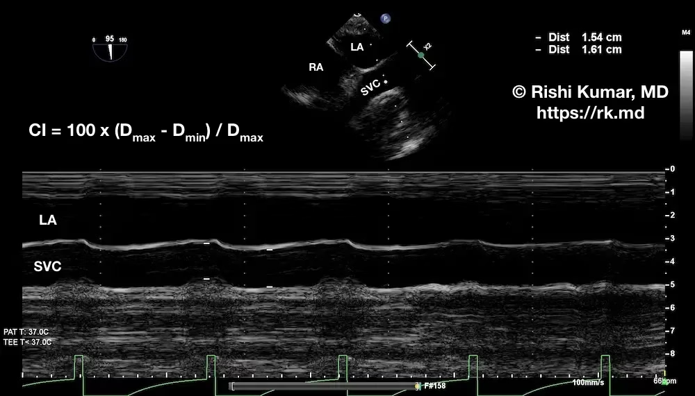
    To determine the superior vena cava (SVC) collapsibility index (CI), I use transesophageal echocardiography (TEE) to obtain a midesophageal bicaval view of the SVC in the long axis. I’ll then apply M-mode through the SVC to determine the maximum and minimum diameters (Dmax and Dmin, respectively) over the respiratory cycle.…

    Read More

  • POCUS – Type B Aortic Dissection

    POCUS – Type B Aortic Dissection

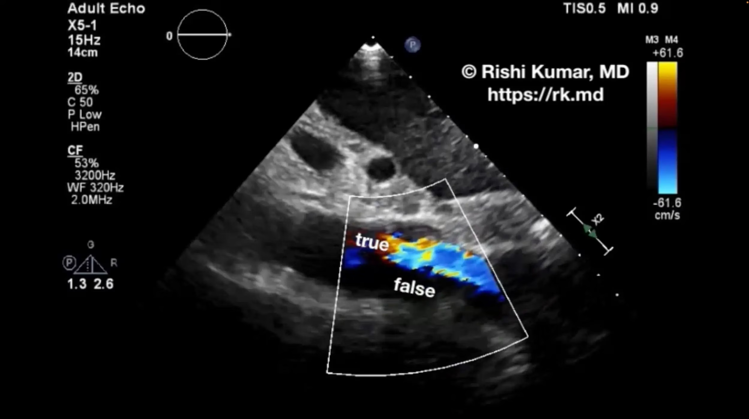
    Point-of-care ultrasound (POCUS) can be a quick and noninvasive modality to confirm a concerning pathology like type B aortic dissection. As a CVICU intensivist, I often receive transfers for “aortic dissection” from other hospitals based on suboptimal imaging. However, they’re sometimes found to be other acute aortic syndromes like intramural…

    Read More

  • Mitral Annular Calcification (MAC)

    Mitral Annular Calcification (MAC)

    Mitral annular calcification (MAC) is a degenerative process that affects the fibrous ring around the mitral valve. It is thought to be caused by chronic inflammation, oxidative stress, and altered calcium metabolism. Although often asymptomatic, MAC can cause mitral valve stenosis and/or regurgitation, with associated symptoms ranging from shortness of…

    Read More

  • Subclavian Vein Cannulation With Ultrasound

    Subclavian Vein Cannulation With Ultrasound

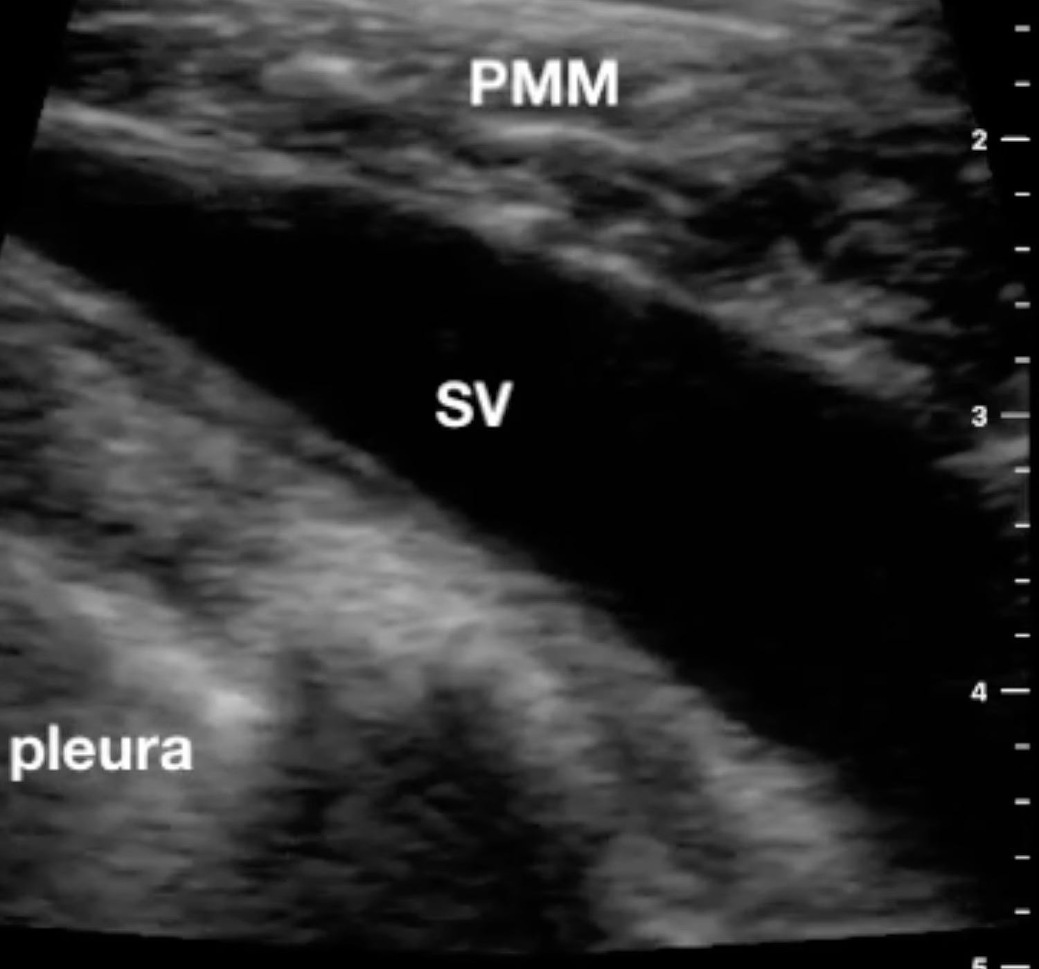
    I’ve performed countless subclavian central line placements using anatomic landmarks; however, more recently, I’m preferentially utilizing ultrasound to cannula the vein from an infraclavicular approach. Because of the proximity of the clavicle to the subclavian artery and vein, I start laterally to locate the axillary artery and vein short axis.…

    Read More

  • Suprasternal Aortic Arch Ultrasound View

    Suprasternal Aortic Arch Ultrasound View

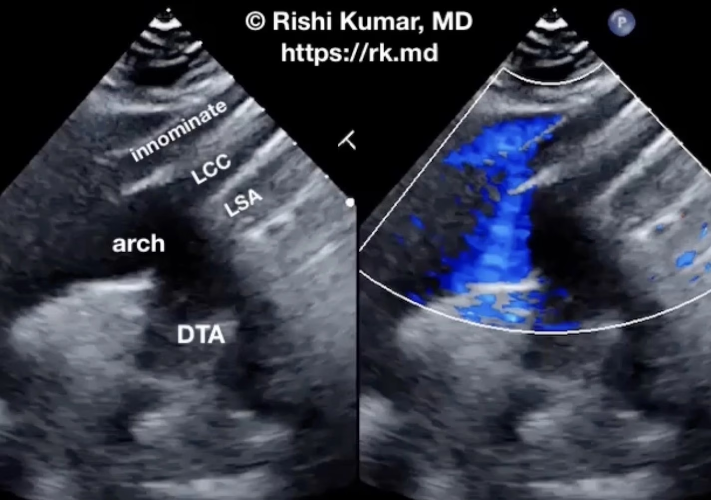
    Suprasternal ultrasound imaging of the aortic arch permits visualization of the great vessels in addition to the aortic arch’s diameter, presence of pathology (e.g., aneurysm, dissection flap), and retrograde flow as seen with aortic valve insufficiency. After extending the patient’s neck, I’ll place a phased-array ultrasound probe in the suprasternal…

    Read More

  • Determining Gastric Volume With Ultrasound

    Determining Gastric Volume With Ultrasound

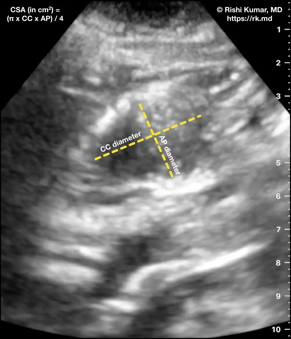
    Gastric ultrasound is a non-invasive, fast, bedside imaging technique that can estimate gastric volume. Placing a curvilinear ultrasound probe (oriented cephalad-caudad) in the epigastric region shows the gastric antrum (A) adjacent to the left lobe of the liver and superficial to the superior mesenteric artery (SMA), abdominal aorta (Ao), and…

    Read More

  • Sulfur Hexafluoride

    Sulfur Hexafluoride

    Sulfur hexafluoride (Lumason) is a medical contrast agent used for ultrasound imaging. It is a clear, colorless, and odorless liquid that is injected into the bloodstream, where it forms tiny bubbles that reflect ultrasound waves. This allows for enhanced visualization of blood vessels, organs, and other structures within the body.…

    Read More

  • Libman-Sacks Endocarditis

    Libman-Sacks Endocarditis

    Libman-Sacks endocarditis (LSE) is a nonbacterial endocarditis associated with lupus, malignancy, and antiphospholipid antibody syndrome characterized by endothelial injury and immune complex deposition typically on left-heart valves (mitral > aortic) causing inflammation and scarring. Unfortunately, symptoms are usually related to embolic sequelae such as stroke, abdominal pain related to gut…

    Read More