
Anatomically, the trachea extends from the cricoid cartilage (level of C6/C7) to the carina (T4/T5) and is reinforced anteriorly by approximately 20 semicircular, cartilaginous rings. The trachea divides into the right and left mainstem bronchi which further divide into secondary bronchi (RUL, RML, RLL, LUL, and LLL). Of note, the bronchus intermedius (BI) is a continuation of the right mainstem bronchus distally. In parentheses, I also included Boyden’s segmental bronchial anatomy nomenclature.
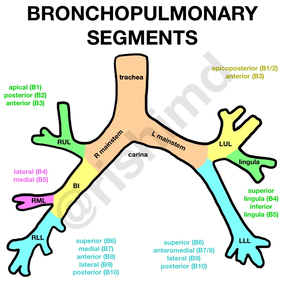
Each of the secondary bronchi give rise to several bronchopulmonary segments – the functional anatomic unit of the lung parenchyma. The right and left lung each have ten of these segments which, in turn, each have a tertiary bronchus, pulmonary artery, and bronchial artery supply. These segments can be identified radiographically and often constitute the boundaries for surgical resection.
It’s important to understand this anatomy during bronchoscopy and airway instrumentation, so we know exactly where we are in the tracheobronchial tree and can communicate the location of our findings more precisely with colleagues.
Remember that the RLL’s posterior segment is a common site for aspiration in the standing position whereas the RLL’s superior segment is more common in the supine position.
Drop me a comment below with questions! 🙂


I ❤️ you Rishi! I could’ve really used this, and you when I was in school. I love the way you’ve explained it. Makes perfect sense. Simply put, and perfection ????
Thanks so much for the sweet comment, Sharon! Glad it helped! 🙂