
There’s a complex yet beautiful architecture within the heart’s fibrous skeleton and valvular geometry. To become a proficient perioperative echocardiographer, one must understand these spatial relationships in three dimensions and the implications that specific surgeries can have on multiple anatomic regions.
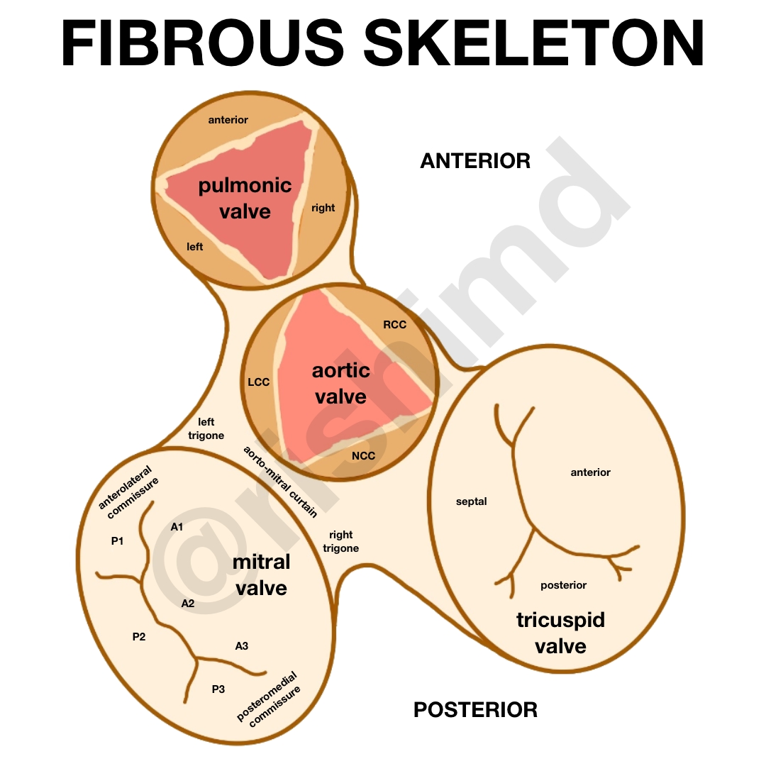
Dense connective tissue forms a fibrous scaffold between the cardiac atria and ventricles to provide electrical insulation, a foundation for attaching the myocardium, and anchors for the valves (namely the tricuspid annulus and mitral annulus). The four cardiac valves are located at different angles and heights relative to each other, so knowing how to best image each valve’s segments is a difficult yet essential part of my training in cardiothoracic anesthesiology.
Drop me a comment below with questions! 🙂


This is a great diagram and explanation. Thank you for sharing.
Sharon Hansen
You’re very welcome, Sharon! Thanks for the comment! 🙂