
In this ultrasound still image, I have a phase array probe positioned on the left side of the body around the 8th rib. A left-sided pleural effusion is noted. It’s important to appreciate this view to look for free fluid in the subphrenic and splenorenal spaces. The green arrow denotes my targeted approach under realtime ultrasound to perform a thoracentesis (drainage of the pleural effusion).
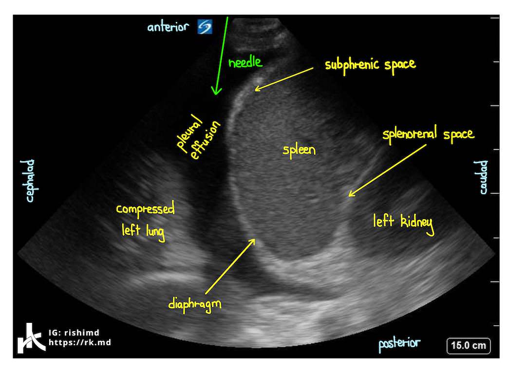
Now let’s look at this same image in motion. The ultrasound transducer is transmitting sound waves from anterior to posterior (top to bottom in the image). Typically, air-filled lungs greatly attenuate the ultrasound beams returning from the spine and posterior chest wall; however, consolidation, effusion, atelectasis, pneumonia and other pathologies provide a denser medium for sound waves to traverse. In this case, reflected waves are able to return to the transducer through the pleural effusion with less distortion thereby allowing us to visualize the vertebral column – the so called “spine sign.” Keep in mind that this is abnormal.

Oh, and what’s that flapping around in the effusion? Compressed lung!
It’s amazing how useful bedside ultrasound is for diagnostics and guiding therapeutic interventions! Drop me a comment below with questions! 🙂


