
In order for the heart muscle to actually contract, the physiological process of excitation-contraction coupling must occur via an action potential propagated through the myocardium. Normally, these muscle cells have a resting transmembrane potential (TMP) around -90 mV (more negative intracellularly than extracellularly). This gradient is created and changed by a flux of ions via concentration/electrical gradients and membrane permeability.
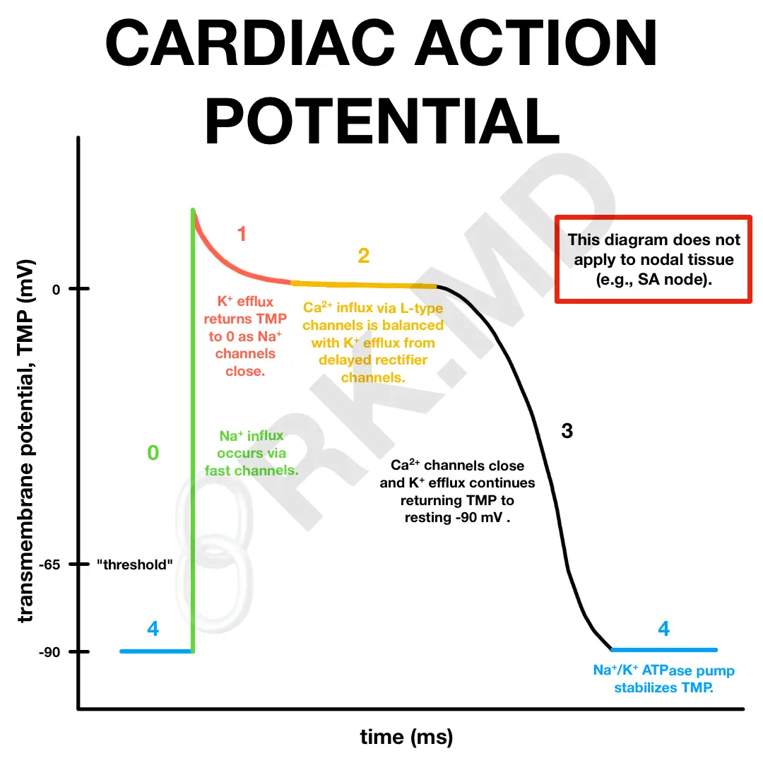
During depolarization, a series of steps occurs:
- Phase 0: An influx of sodium (Na+) via fast channels begins increasing the TMP and actually overshoots to around +10 mV.
- Phase 1: An initial repolarization via efflux of K+ out of the cell brings the TMP back to 0 mV.
- Phase 2: Ca2+ influx is balanced with K+ efflux causing a plateau.
- Phase 3: As the Ca2+ channels close and K+ efflux continues, the TMP is brought back to its resting potential.
- Phase 4: Resting potential is maintained via a Na+/K+ ATPase pump.
By understanding these channels, it makes sense how medications like digoxin (myocardial Na+/K+ ATPase pump inhibitor), sodium channel blocking antiarrhythmics, and calcium channel blockers can provide therapeutic benefits. Keep in mind that this diagram does NOT describe nodal tissues (like the SA node) which contain slightly different channels to achieve automaticity.
Drop me a comment below with questions!


How do these action potentials differ from cardiac pacemaker action potentials? At what point do HCN channels become active in the pacemaker cells and do they work in conjunction with the Na/K ATPase, or do they replace their function?