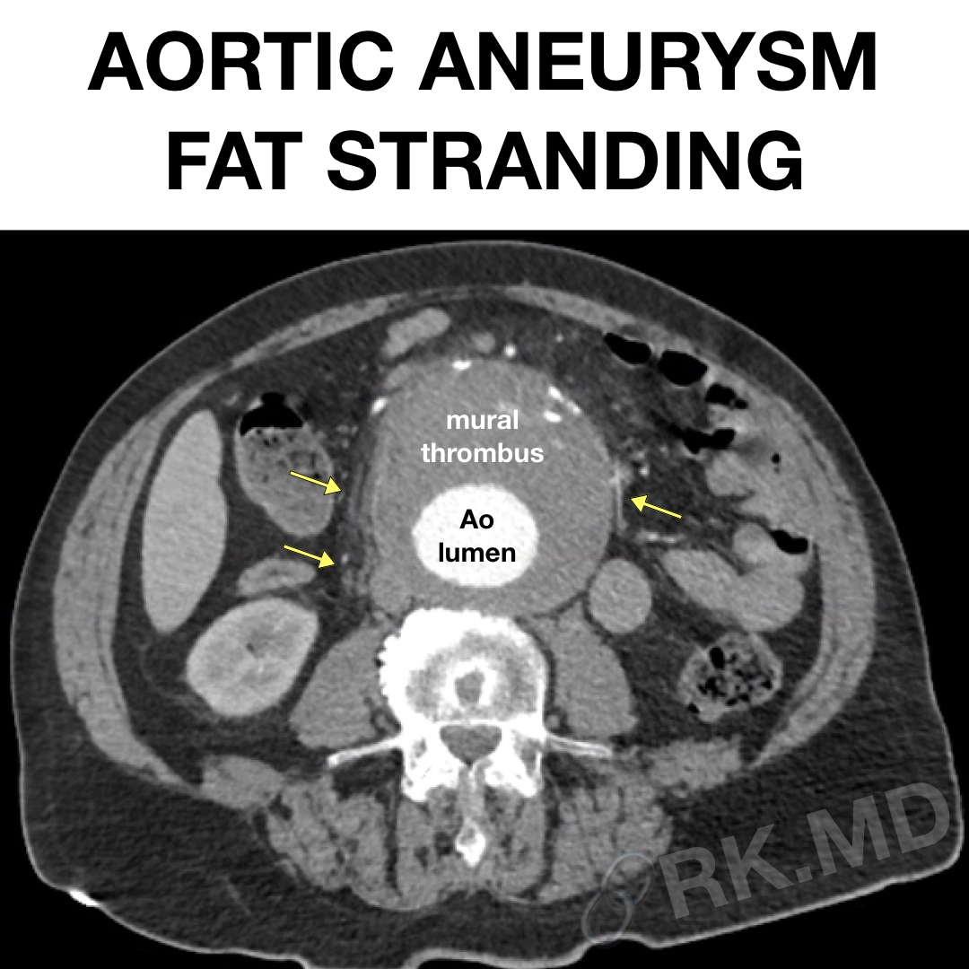
As a cardiac anesthesiologist and cardiovascular intensivist, I care for many patients with aortic aneurysms in the perioperative setting. Contrast-enhanced CT imaging is traditionally utilized to assess the location and size of the aneurysm and its influence on surrounding structures like compressive mass effect, fistulas, dissection, etc.
If the aneurysm becomes large enough, it can pressure nearby organs and tissues, including the fatty tissue surrounding the aorta. This pressure can cause inflammation and swelling in the fatty tissue, known as fat stranding.
In the pictured transverse and coronal CT images of a large abdominal aortic aneurysm, the yellow arrows indicate areas of fat stranding. Although there is no evident extravasation of contrast from the aortic (Ao) lumen, periaortic fat stranding may be a sign of pressure on the surrounding tissue or a possible impending rupture of the aneurysm. This necessitates an emergent intervention such as an endovascular stent graft placement.


