
Boerhaave syndrome, first described by the Dutch physician Hermann Boerhaave in 1724, is a life-threatening condition involving spontaneous esophageal perforation from a sudden, forceful increase in intraesophageal pressure against a closed glottis (e.g., intense vomiting, retching, or straining). The most common perforation site is the esophagus’s distal third, particularly along the left posterolateral wall. The mediastinum becomes vulnerable to contamination by gastric contents, leading to mediastinitis – a rapidly progressive and often fatal infection without intervention.
Boerhaave syndrome often presents with Mackler’s triad: severe chest pain, vomiting, and subcutaneous emphysema. Other symptoms include dyspnea, fever, tachycardia, or hemodynamic instability as the condition progresses. A chest X-ray may reveal pneumomediastinum, pleural effusion (often left-sided), or subcutaneous emphysema. A CT scan with oral contrast or an esophagogram may show contrast extravasation.
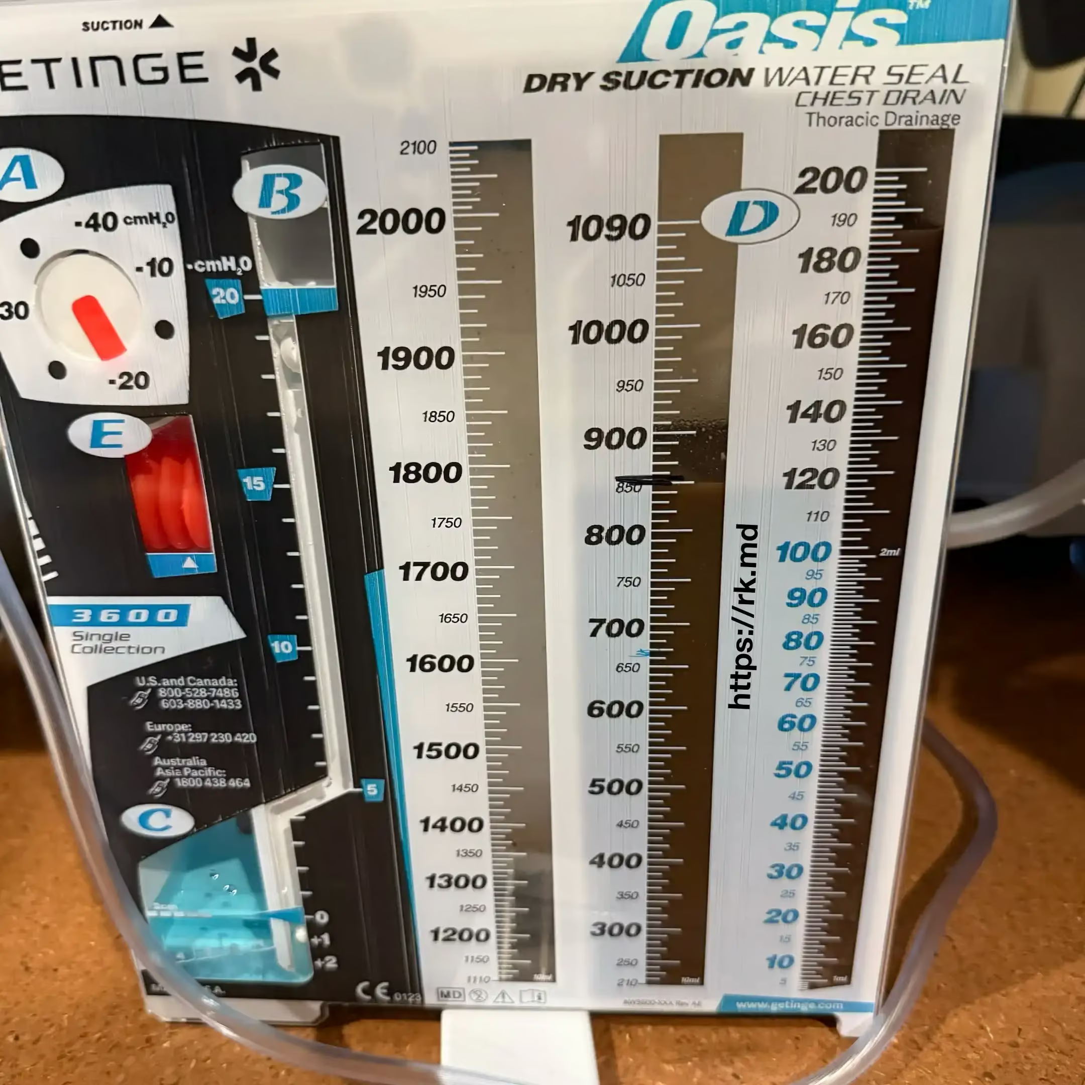
Management is largely centered around the location, size, and timing of the diagnosis and involves resuscitation, broad-spectrum antibiotics to cover polymicrobial flora, NPO, and source control of the detected (surgical repair, stenting, drainage of the mediastinal/pleural fluid collections, etc.).
This syndrome demonstrates how even physiologic events like vomiting can lead to catastrophic complications requiring a high index of suspicion, multidisciplinary management, and aggressive therapy to reduce morbidity and mortality!


Leave a Reply