• Lung Point For Pneumothorax Using Ultrasound

    Lung Point For Pneumothorax Using Ultrasound

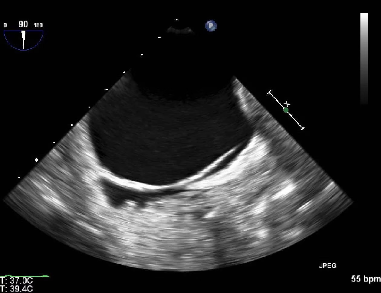
    With a pneumothorax (PTX), air separates the visceral and parietal pleura layers located on the lung’s outside surface and inner chest wall, respectively. The absence of pleural sliding on ultrasonography can suggest PTX, but when this is found adjacent to a lung point, it’s 100% specific for a PTX. The…

    Read More

  • Alprostadil

    Alprostadil

    Alprostadil (Prostaglandin E1, PGE1) is a medication used to treat erectile dysfunction and pulmonary arterial hypertension (PAH). PAH is a progressive condition in which the arteries that carry blood from the heart to the lungs become narrowed, making it difficult for the heart to pump blood effectively. This can lead…

    Read More

  • Aortic Aneurysm Fat Stranding On CT

    Aortic Aneurysm Fat Stranding On CT

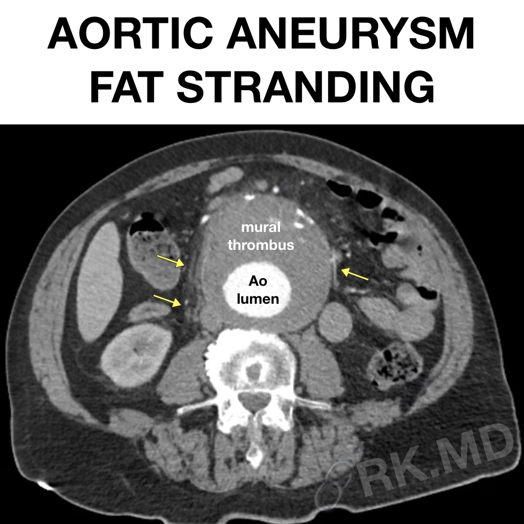
    As a cardiac anesthesiologist and cardiovascular intensivist, I care for many patients with aortic aneurysms in the perioperative setting. Contrast-enhanced CT imaging is traditionally utilized to assess the location and size of the aneurysm and its influence on surrounding structures like compressive mass effect, fistulas, dissection, etc. If the aneurysm…

    Read More

  • Break Down Intake And Output (I&Os)

    Break Down Intake And Output (I&Os)

    As a cardiac anesthesiologist and cardiovascular intensivist, I routinely care for perioperative cardiac surgery patients in my OR and ICU practices. If I were to look at the “I&Os” (intake and output) to estimate how “net positive” a patient is for their hospital admission without breaking down the types of…

    Read More

  • D-Sign: Right Ventricular Strain On Chest CT And Echocardiography

    D-Sign: Right Ventricular Strain On Chest CT And Echocardiography

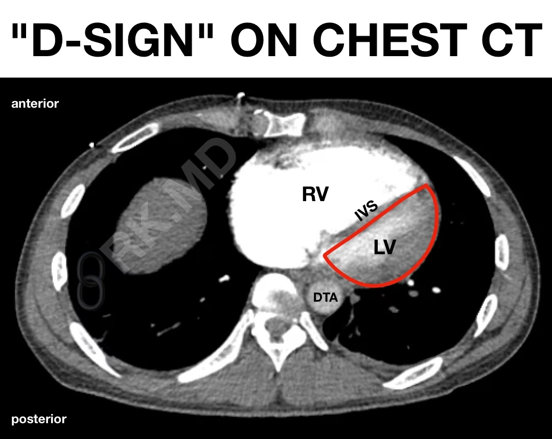
    Typically at end-systole, the interventricular septum (IVS) bows rightward as the left ventricle’s pressure (LV) exceeds that of the right ventricle (RV). However, when the RV is strained (e.g., massive pulmonary embolism), the pressure overload can lead to flattening or even paradoxical bowing of the IVS. In a short-axis view…

    Read More

  • Fluorescein

    Fluorescein

    Fluorescein is a dye that is used in a variety of medical and scientific applications. For example, it is often used as a diagnostic tool to help identify and treat eye conditions and visualize blood flow in the body. In the eye, fluorescein detects and diagnoses conditions such as corneal…

    Read More

  • Bivalirudin

    Bivalirudin

    Bivalirudin (Angiomax) is an intravenous direct thrombin inhibitor used as an anticoagulant for acute coronary syndrome, mechanical circulatory support, and cardiopulmonary bypass when patients have a contraindication to use heparin (e.g., heparin-induced thrombocytopenia). The usual dose of bivalirudin is a bolus of 0.75 mg/kg followed by an infusion of 1.75…

    Read More

  • My Transesophageal Echocardiography (TEE) Sequence Of Views

    My Transesophageal Echocardiography (TEE) Sequence Of Views

    As a cardiothoracic anesthesiologist and intensivist, transesophageal echocardiography (TEE) is a powerful cardiac imaging modality I perform in the OR during cardiac surgery and in the cardiovascular ICU when point-of-care ultrasound is not feasible or helpful due to poor image quality. Before anything, I take a quick shot of the…

    Read More

  • TAPSE With TEE

    TAPSE With TEE

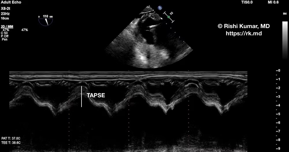
    Tricuspid annular plane systolic excursion (TAPSE) is a quantitative echocardiography technique to assess right ventricular (RV) systolic function. As an intensivist, I obtain an apical four-chamber view using transthoracic echocardiography, activate motion (“M”) mode through the lateral tricuspid valve (TV) annulus, and measure the peak-to-trough distance. A value > 18…

    Read More

  • Hypochloremic (“Contraction”) Alkalosis

    Hypochloremic (“Contraction”) Alkalosis

    Metabolic alkalosis is a derangement characterized by increased plasma bicarbonate (HCO3–), resulting in a pH > ~7.45. This can be a product of hydrogen ion (H+) loss (e.g., vomiting or renal losses), intracellular shifting of H+, or retention/creation of HCO3–. Typically, the proximal convoluted tubule’s (PCT) ion channels reabsorb all…

    Read More