• Saddle Pulmonary Embolism (PE)

    Saddle Pulmonary Embolism (PE)

    Saddle pulmonary embolism (SPE) is a severe and life-threatening condition when a blood clot (embolus) becomes lodged in the main pulmonary artery, blocking blood flow to both lungs. The term “saddle” refers to the shape of the clot, which straddles the bifurcation of the pulmonary artery, resembling a saddle. The…

    Read More

  • Physician Modified Endovascular Graft (PMEG)

    Physician Modified Endovascular Graft (PMEG)

    Physician-modified endovascular grafts (PMEGs) offer a percutaneous and effective option for repairing complex thoracoabdominal aortic aneurysms. A stent graft is modified onsite using strict sterile technique with reinforced fenestrations created based on the patient’s specific anatomy. Additionally, radiopaque markers are added to help with stent graft alignment in vivo. In…

    Read More

  • Suicide Left Ventricle Following TAVR

    Suicide Left Ventricle Following TAVR

    Calcific aortic valve stenosis causes the left ventricle (LV) to adapt to chronic pressure overload over time. Classically, this involves left ventricular hypertrophy resulting in diastolic dysfunction. After a transcatheter aortic valve replacement (TAVR), the new valve instantaneously alleviates the fixed obstruction. As a result, the LV can eject more…

    Read More

  • Subclavian Vein Cannulation With Ultrasound

    Subclavian Vein Cannulation With Ultrasound

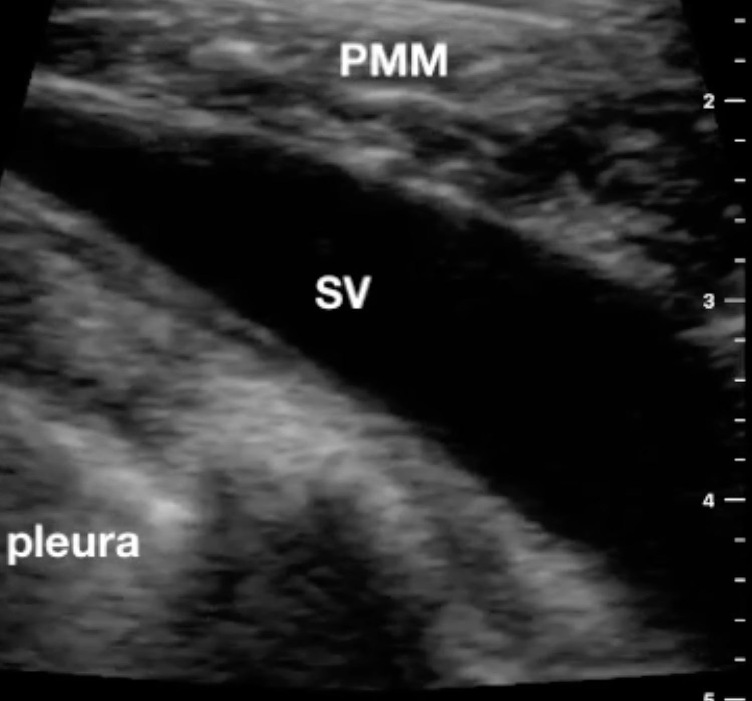
    I’ve performed countless subclavian central line placements using anatomic landmarks; however, more recently, I’m preferentially utilizing ultrasound to cannula the vein from an infraclavicular approach. Because of the proximity of the clavicle to the subclavian artery and vein, I start laterally to locate the axillary artery and vein short axis.…

    Read More

  • Fosphenytoin

    Fosphenytoin

    Fosphenytoin (Cerebyx) is a prodrug of phenytoin, an antiepileptic drug used to treat seizures. Unlike phenytoin, which is poorly soluble in water and can cause severe local irritation if injected into the muscle, fosphenytoin is water-soluble and can be administered intravenously without causing tissue damage. Fosphenytoin is rapidly converted to…

    Read More

  • Suprasternal Aortic Arch Ultrasound View

    Suprasternal Aortic Arch Ultrasound View

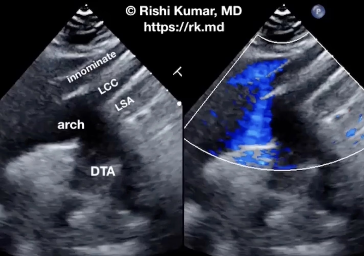
    Suprasternal ultrasound imaging of the aortic arch permits visualization of the great vessels in addition to the aortic arch’s diameter, presence of pathology (e.g., aneurysm, dissection flap), and retrograde flow as seen with aortic valve insufficiency. After extending the patient’s neck, I’ll place a phased-array ultrasound probe in the suprasternal…

    Read More

  • Determining Gastric Volume With Ultrasound

    Determining Gastric Volume With Ultrasound

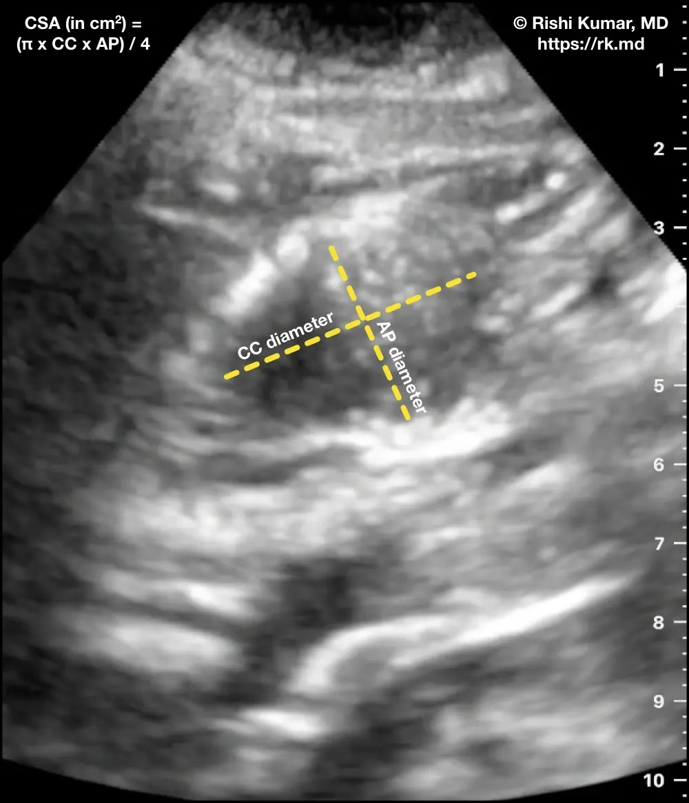
    Gastric ultrasound is a non-invasive, fast, bedside imaging technique that can estimate gastric volume. Placing a curvilinear ultrasound probe (oriented cephalad-caudad) in the epigastric region shows the gastric antrum (A) adjacent to the left lobe of the liver and superficial to the superior mesenteric artery (SMA), abdominal aorta (Ao), and…

    Read More

  • Sulfur Hexafluoride

    Sulfur Hexafluoride

    Sulfur hexafluoride (Lumason) is a medical contrast agent used for ultrasound imaging. It is a clear, colorless, and odorless liquid that is injected into the bloodstream, where it forms tiny bubbles that reflect ultrasound waves. This allows for enhanced visualization of blood vessels, organs, and other structures within the body.…

    Read More

  • Libman-Sacks Endocarditis

    Libman-Sacks Endocarditis

    Libman-Sacks endocarditis (LSE) is a nonbacterial endocarditis associated with lupus, malignancy, and antiphospholipid antibody syndrome characterized by endothelial injury and immune complex deposition typically on left-heart valves (mitral > aortic) causing inflammation and scarring. Unfortunately, symptoms are usually related to embolic sequelae such as stroke, abdominal pain related to gut…

    Read More

  • Double Aortic Arch

    Double Aortic Arch

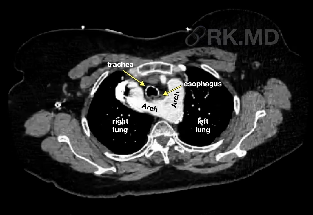
    During embryogenesis, the aortic sac is connected to paired dorsal aortae via six symmetrical aortic (Ao) arches, which give rise to major vascular structures. For example, the right and left common carotid arteries originate from the right and left third arches, respectively. The proximal right subclavian artery is formed from…

    Read More