Hydromorphone
My opioid of choice in the perioperative and ICU settings is hydromorphone (Dilaudid). This medication is roughly 5-10 times more potent than morphine and comes in intravenous (IV) and oral formulations.


My opioid of choice in the perioperative and ICU settings is hydromorphone (Dilaudid). This medication is roughly 5-10 times more potent than morphine and comes in intravenous (IV) and oral formulations.

Remifentanil (Ultiva) is a potent (~200-300 times more than morphine), short acting, titratable narcotic infusion I use for moderate/deep sedation and maintaining general anesthetics. Unlike many drugs which rely on hepatic and/or renal metabolism, remifentanil’s metabolism is through nonspecific plasma esterases. This explains why the drug does not “build up”…
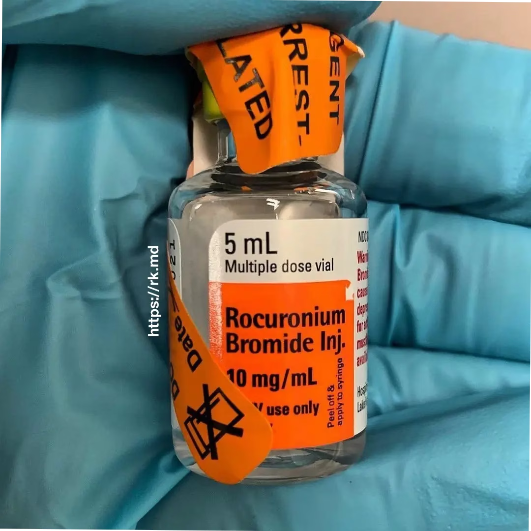
Rocuronium is an aminosteroid nondepolarizing neuromuscular junction blocker (NMJB) I routinely use to facilitate tracheal intubation. The name rocuronium comes from the fact that it’s a rapid onset -curonium (referring to its steroid structure). With a “rapid sequence” dose (usually 1.2 mg/kg), it has an onset almost as fast as succinylcholine without…

The continuity equation illustrates the conservation of energy. In the case of blood flow within the (normal) heart, volumetric flow (cardiac output) at serial sites should be equal. The image shows flow velocities across the left ventricular outflow tract (LVOT) and through the aortic valve. Because these structures are in…

The Doppler effect refers to a shift in frequency perceived by an object in motion relative to the source of a wave. When applying this to ultrasonography, the transducer is a stationary source of ultrasound waves of a known frequency (depending on the mode) and velocity (1540 meters/second) which reflect…
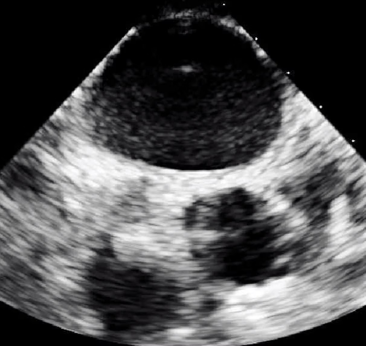
Today’s didactic lecture on epiaortic ultrasonography was given by one of my wonderful cardiothoracic anesthesiology attendings – Dr. Jeff Swanson. As part of traditional cardiac surgeries, the aorta can be extensively manipulated – especially when performing arterial cannulation and applying the aortic cross-clamp. This can dislodge plaques systemically leading to…

There’s a complex yet beautiful architecture within the heart’s fibrous skeleton and valvular geometry. To become a proficient perioperative echocardiographer, one must understand these spatial relationships in three dimensions and the implications that specific surgeries can have on multiple anatomic regions.
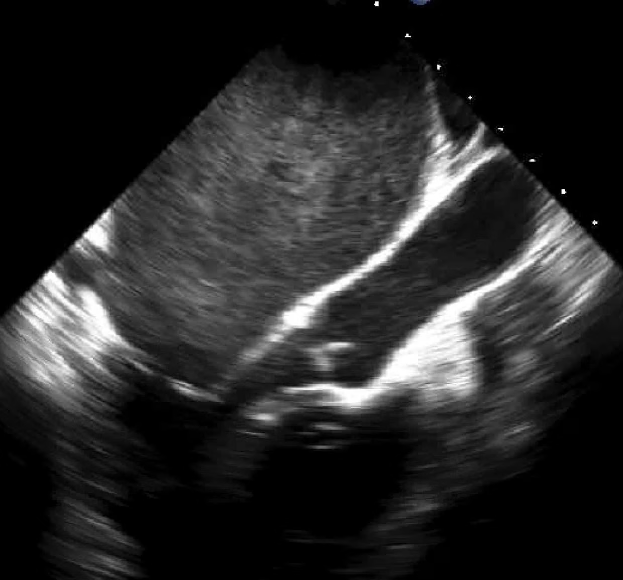
Left atrial spontaneous echo contrast (SEC) is an echogenic, smoke-like swirling of aggregated red blood cells suggestive of stasis. This sign confers an increased risk of thrombosis in the left atrium and/or left atrial appendage. We most commonly see SEC on transesophageal echocardiography (TEE) in patients with atrial arrhythmias like…

Most organ systems have some form of autoregulation to provide a steady supply of blood flow and oxygen to maintain normal physiologic function. This autoregulation is largely driven by chemical mediators to “couple” metabolic demand with blood supply. In other words, if the organ demands less oxygen, then it will…
