• Catheter Sizing – French Versus Gauge

    Catheter Sizing – French Versus Gauge

    Whether it’s a 1.88 inch long 20 gauge peripheral intravenous (PIV) catheter or a 13.5 French Mahurkar dialysis catheter, healthcare workers are exposed to both the “gauge” and “French” unit systems almost every day. As an anesthesiologist and intensivist who places many kinds of catheters, it’s important for me to…
    Read More

  • ProtekDuo® Veno-Venous Cannula

    ProtekDuo® Veno-Venous Cannula

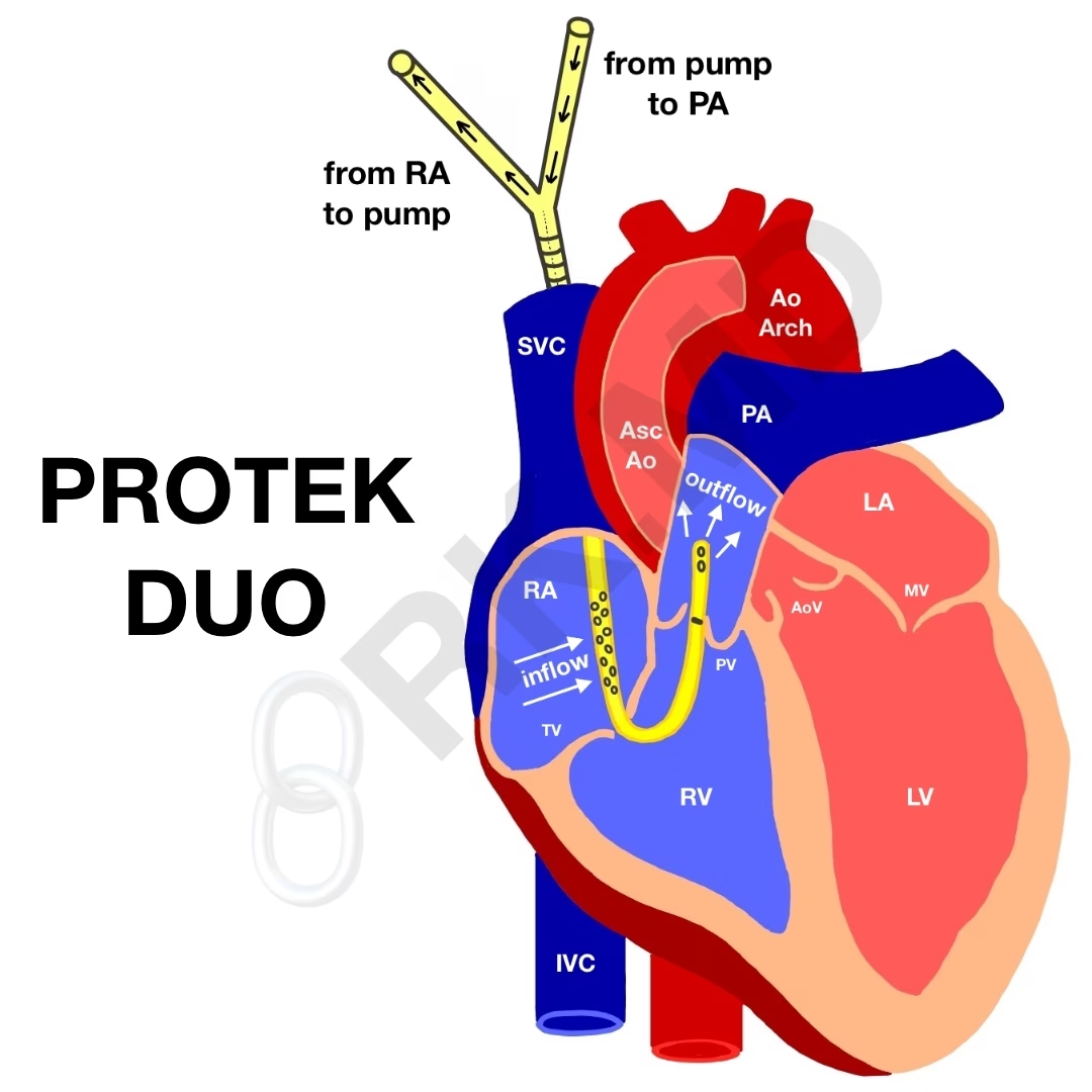
    The ProtekDuo veno-venous (V-V) dual lumen cannula provides extracorporeal mechanical circulatory support (MCS) with or without an oxygenator. When combined with a TandemHeart® pump, it can deliver ~4.5 liters/minute of flow by draining blood from the right atrium and returning it to the pulmonary artery (PA). This allows right side…

    Read More

  • Passy Muir Valve

    Passy Muir Valve

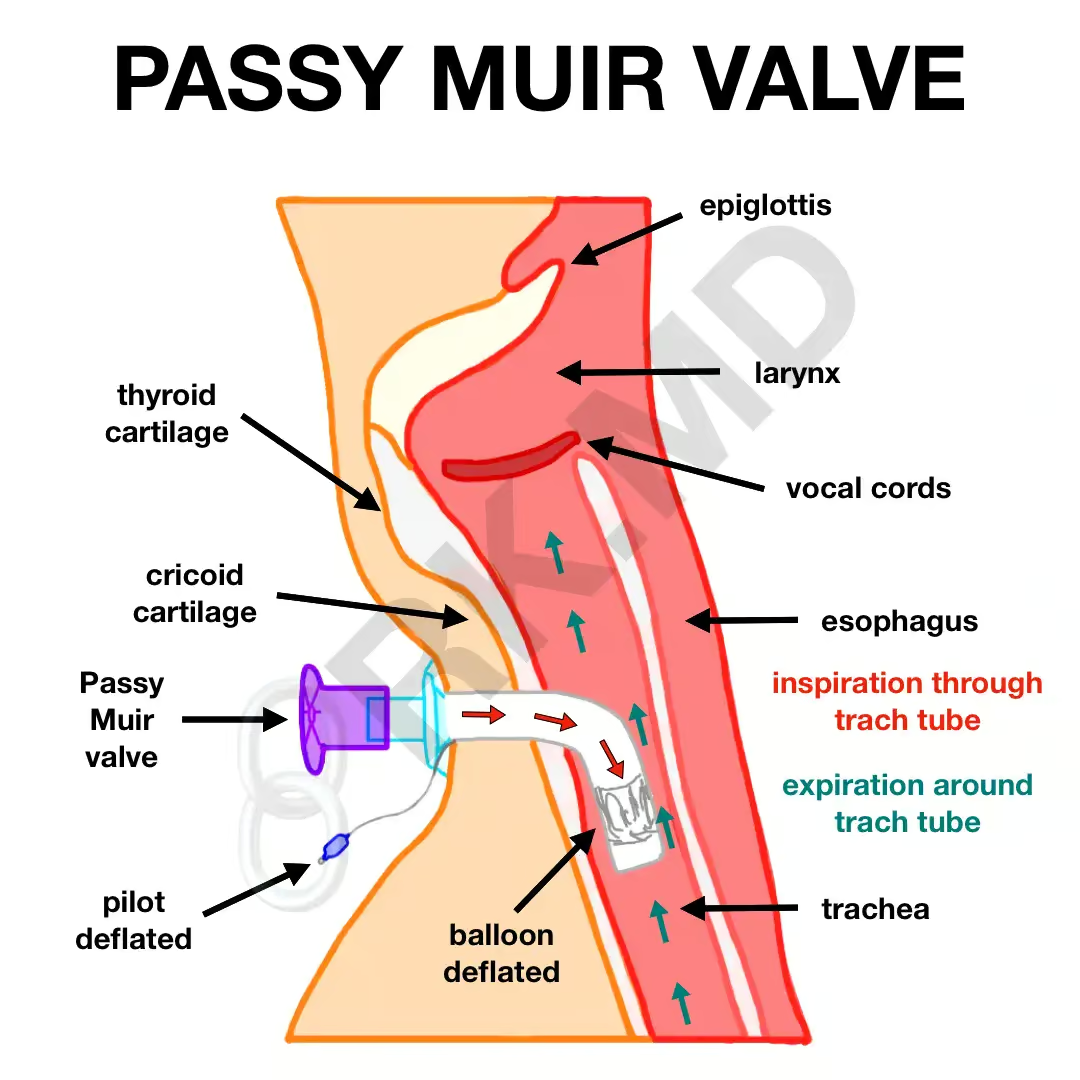
    Normally, air moves in and out of tracheostomy tubes (trachs) below the level of the vocal cords rendering a patient aphonic. However, using a Passy Muir valve (PMV), otherwise known as a “speaking valve,” airflow is redirected through the vocal cords during expiration – our standard mechanism of phonation. This…

    Read More

  • Intracardiac Echocardiography (ICE)

    Intracardiac Echocardiography (ICE)

    Intracardiac echocardiography (ICE) allows cardiologists to visualize cardiac structures (aorta, valves, pulmonary veins, chambers, etc) from an ultrasound catheter usually threaded into the right atrium (RA) or right ventricle (RV) from the femoral vein. It aids in procedures ranging from percutaneous valve replacement and ablation to septal defect closures by…

    Read More

  • Mitral Valve Anatomy

    Mitral Valve Anatomy

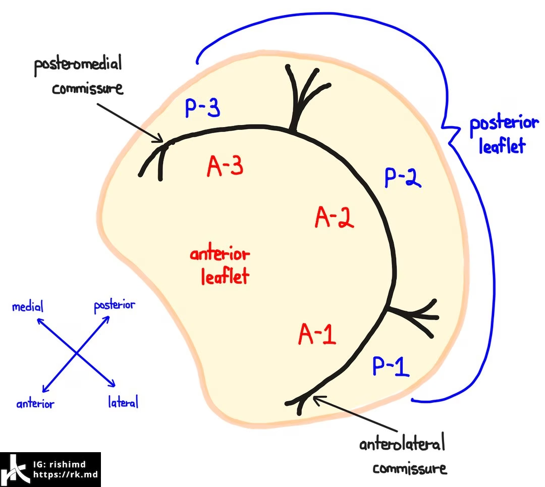
    The mitral valve (MV) separates the left atrium (LA) and left ventricle (LV). It’s a bileaflet valve composed of an anterior and posterior leaflet, each with three “scallops” designated A1, A2, A3, P1, P2, and P3. The A3-P3 interface tapers into the posteromedial commissure whereas the A1-P1 interface becomes the…

    Read More

  • Reexpansion Pulmonary Edema

    Reexpansion Pulmonary Edema

    Reexpansion pulmonary edema (RPE) is a rare but potentially life-threatening condition that occurs when the lung is rapidly expanded following compression due to air (pneumothorax) or fluid (pleural effusion). Although the underlying pathophysiology is poorly understood, it is attributed to increased alveolar-capillary membrane permeability, reperfusion injury, and even surfactant dysfunction.…

    Read More

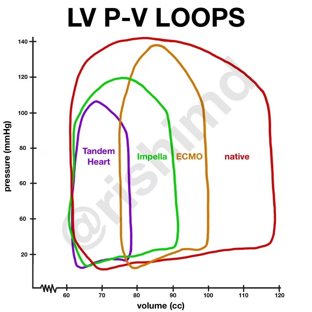
  • Left Ventricular Pressure-Volume Work Loop With Mechanical Circulatory Support
  • Blakemore Tube

    Blakemore Tube

    Blakemore tubes (and their analogues like Minnesota tubes) are rarely used in the modern day of endoscopy but can still have a role in the acute setting of GI variceal bleeding. These tubes are lubricated and passed to roughly 50 centimeters in the same manner as an orogastric or nasogastric…

    Read More

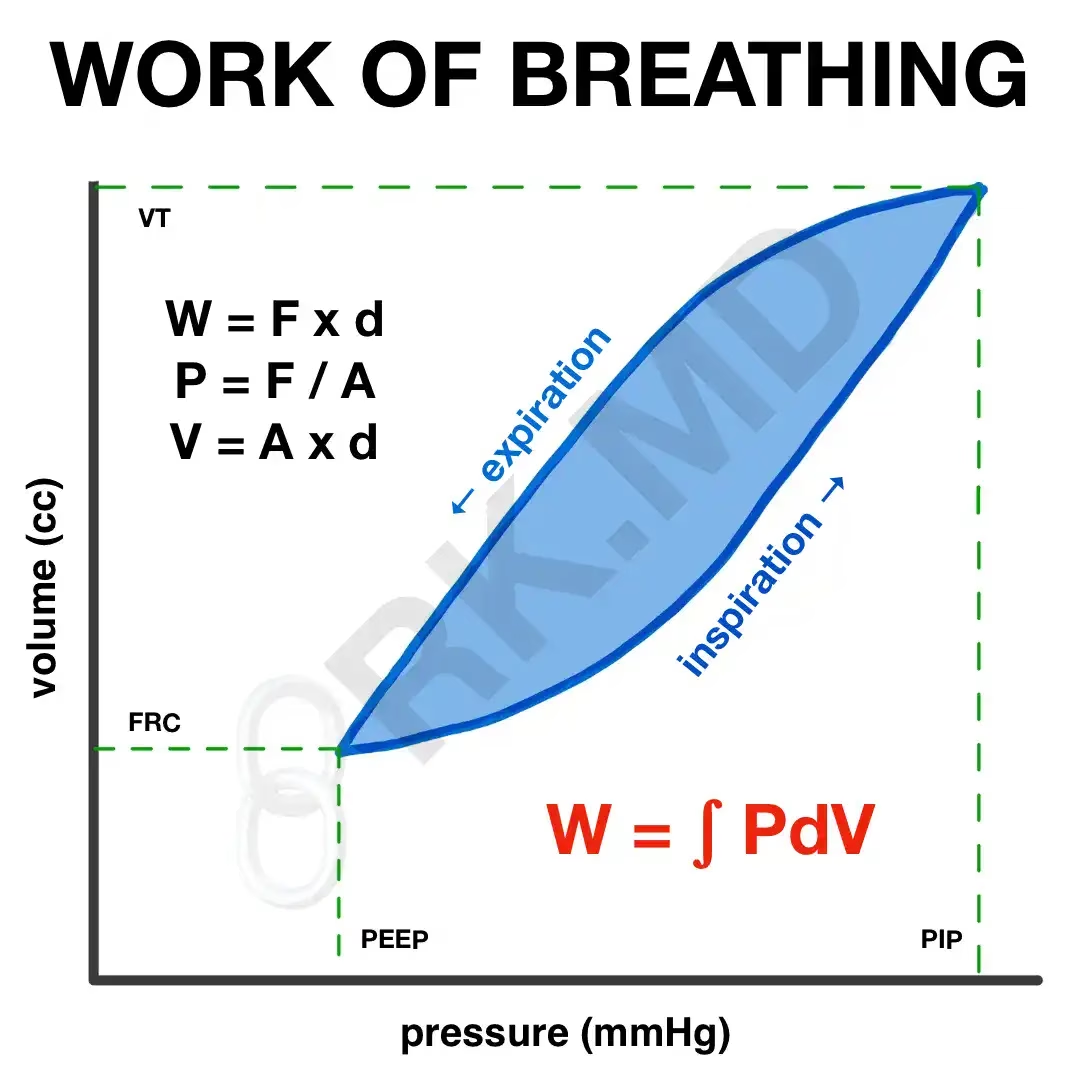
  • Work Of Breathing – Pressure-Volume Curve Integration
  • Pulse Contour Analysis

    Pulse Contour Analysis

    Described over a century ago, pulse contour analysis was purported to be a surrogate for cardiac output. This technique calculates the area under the systolic portion of the arterial pressure curve. This area changes during the respiratory cycle due to changes in intrathoracic pressure and subsequent changes in ventricular preload…

    Read More