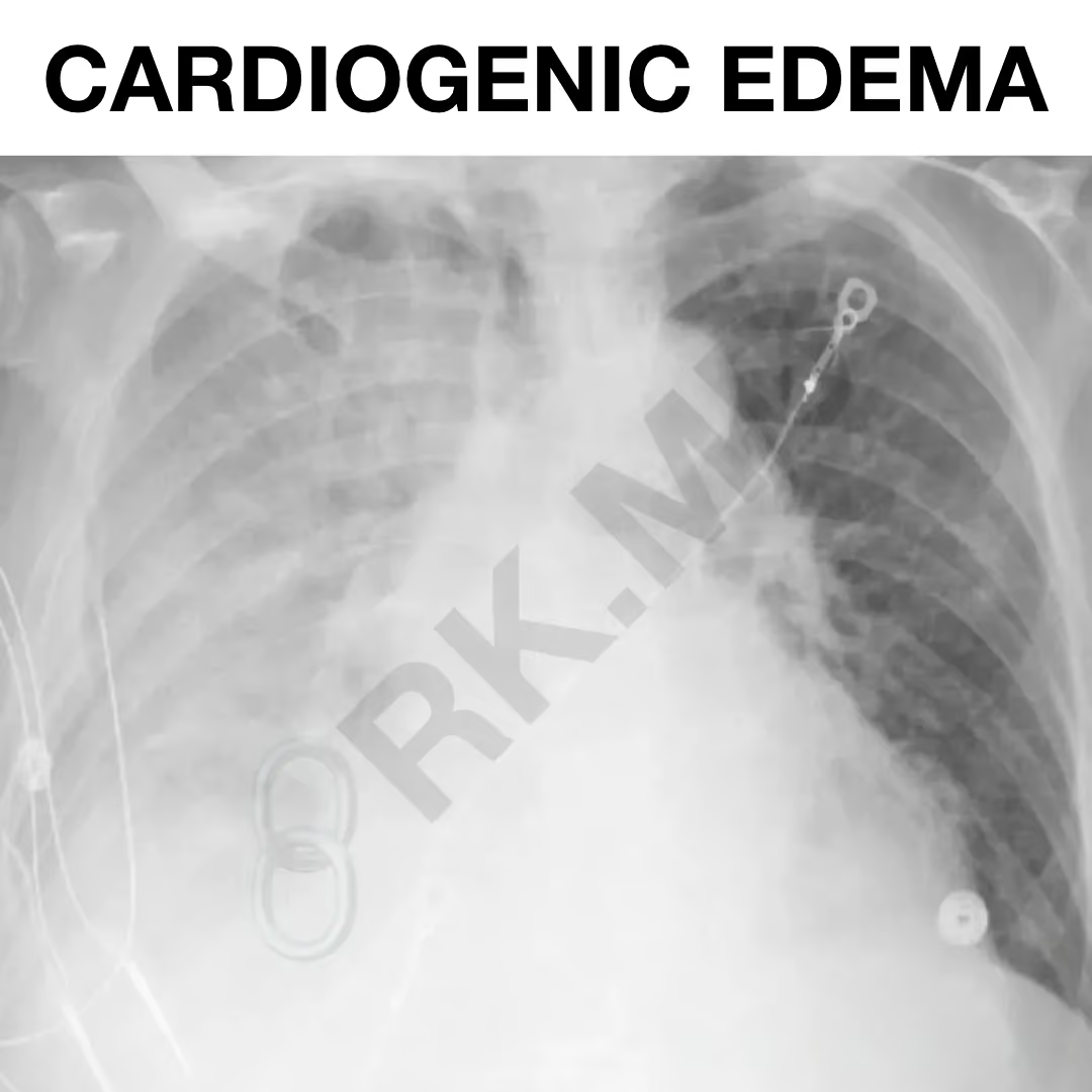
• Unilateral Cardiogenic Pulmonary Edema

    Unilateral Cardiogenic Pulmonary Edema

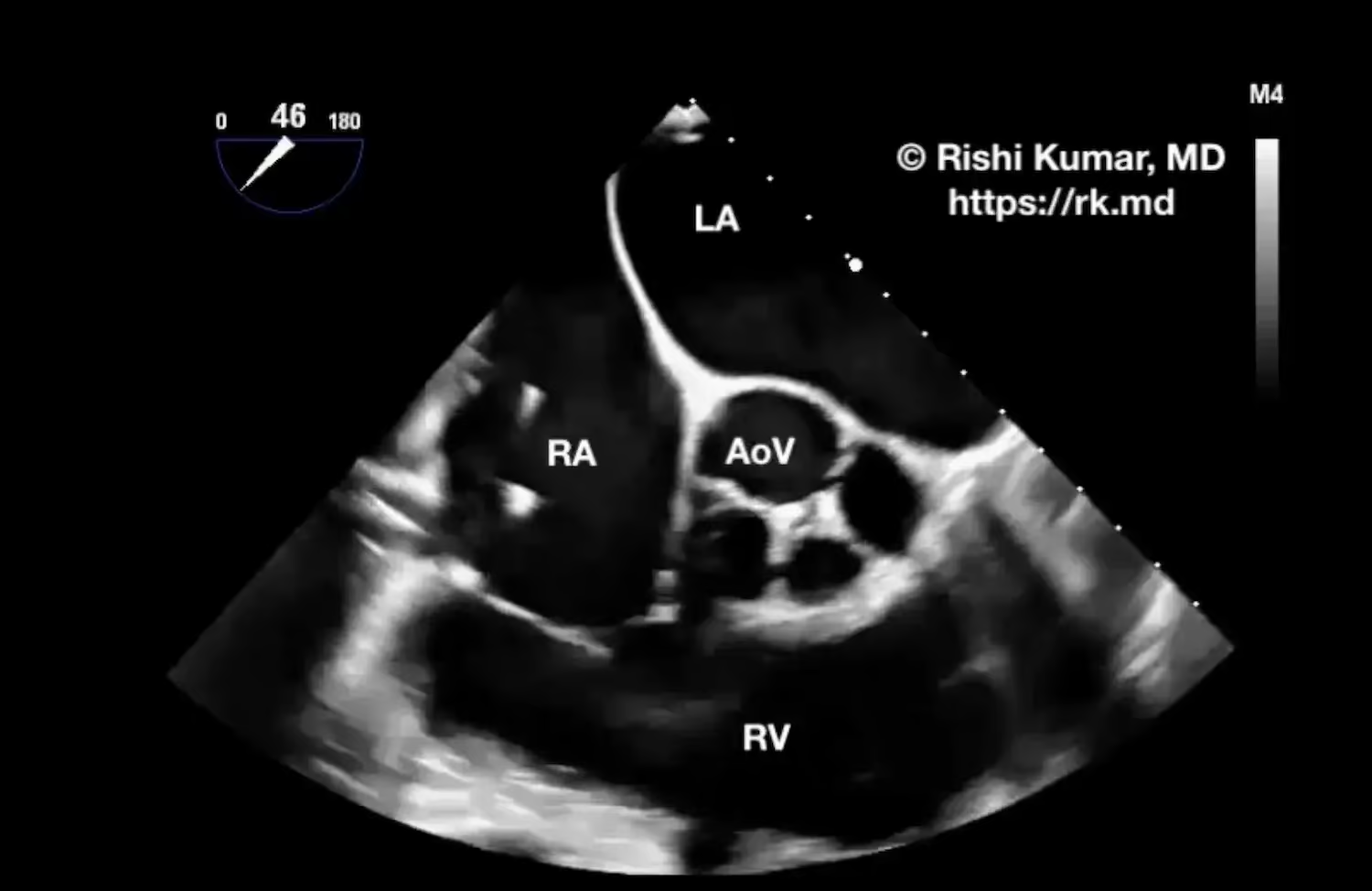
    Unilateral cardiogenic pulmonary edema (CPE) accounts for ~2% of pulmonary edema cases and is classically caused by posterior papillary muscle rupture, resulting in acute, severe, eccentric mitral regurgitation. Flow is directed specifically to the right-sided pulmonary veins. This, in turn, overwhelms the lymphatic capacity of the right lung and results…
    Read More

  • Quadricuspid Aortic Valve

    Quadricuspid Aortic Valve

    The normal aortic valve is trileaflet and comprised of the non, left, and right coronary cusps. Quadricuspid aortic valve (QAV) is an extremely rare malformation occurring in only 0.008% of autopsies and 0.043% of cases examined through echocardiography. Hurwitz and Roberts proposed seven classes of QAV based on the arrangement…

    Read More

  • Aortic Dissection Flap Fenestrations

    Aortic Dissection Flap Fenestrations

    Aortic dissection is a life-threatening condition characterized by a tear in the innermost layer of the aorta (tunica intima), permitting blood to enter and separate the intima from the aorta’s middle layer (tunica media). This event creates a false lumen (FL) alongside the true lumen (TL) of the aorta. Typically,…

    Read More

  • Concentric Left Ventricular Hypertrophy

    Concentric Left Ventricular Hypertrophy

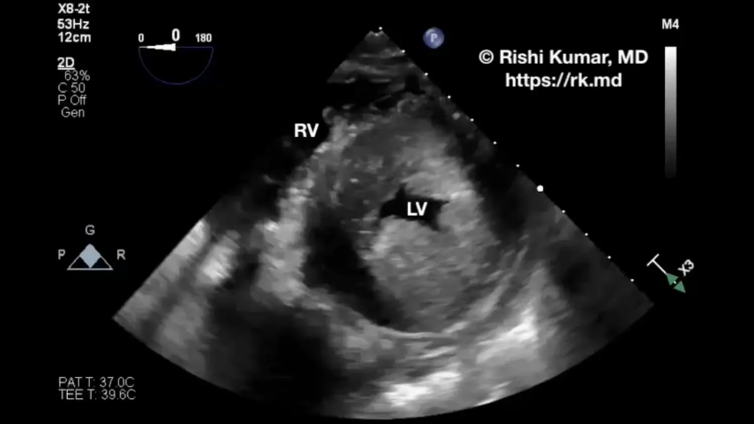
    Concentric left ventricular hypertrophy (LVH) is characterized by adaptive thickening of the myocardium in response to pressure overload from chronic hypertension or aortic valve stenosis. LVH can also be seen in genetic conditions like Fabry disease or hypertrophic cardiomyopathy with chaotic sarcomeric disarray at the cellular level. LVH leads to…

    Read More

  • Estimating Intraabdominal Pressure With Bladder Pressure

    Estimating Intraabdominal Pressure With Bladder Pressure

    Intraabdominal hypertension (IAH) and abdominal compartment syndrome are defined as sustained intraabdominal pressure (IAP) ≥ 12 mmHg and > 20 mmHg with new organ dysfunction, respectively. This can be challenging to diagnose in patients who are pregnant, obese, or with ascites. Bladder pressure from a Foley catheter can be used…

    Read More

  • Pulmonary Velocity Acceleration Time (PVAT)

    Pulmonary Velocity Acceleration Time (PVAT)

    Pulmonary velocity acceleration time (PVAT), measured in milliseconds (ms), can be used to estimate pulmonary artery pressure (PAP) when a tricuspid regurgitation jet is difficult to locate or cannot be accurately interrogated with Doppler. In this parasternal short-axis transthoracic echo view directed anteriorly and to the patient’s right, pulsed-wave Doppler…

    Read More

  • Cardiac Standstill POCUS

    Cardiac Standstill POCUS

    Point-of-care ultrasound (POCUS) can be extremely helpful during advanced cardiovascular life support (ACLS) with ongoing chest compressions and resuscitation. POCUS can help rule in or out certain etiologies (the H’s and T’s) for why a patient arrested. During the pulse check, I record the clearest subcostal 4 or 5 chamber…

    Read More

  • POCUS – Transhepatic IVC View

    POCUS – Transhepatic IVC View

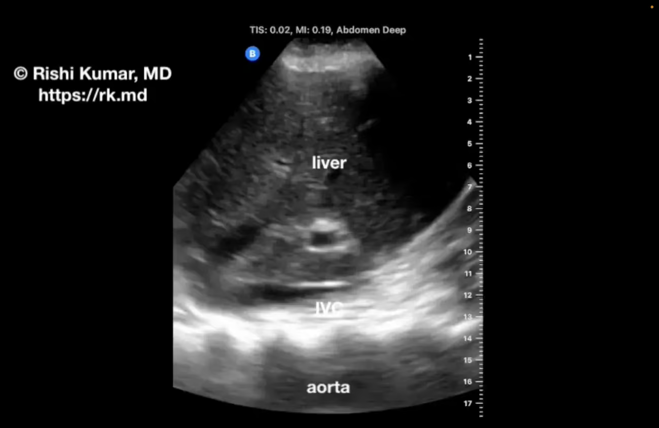
    The traditional way of assessing the inferior vena cava (IVC) with point-of-care ultrasound (POCUS) is longitudinally from a subcostal window; however, this can be challenging when that area is off limits (subcostal chest tubes, burn injuries, etc.) The transhepatic IVC view is obtained by placing a phased array probe (oriented…

    Read More

  • Carpentier Type III Mitral Regurgitation On TEE

    Carpentier Type III Mitral Regurgitation On TEE

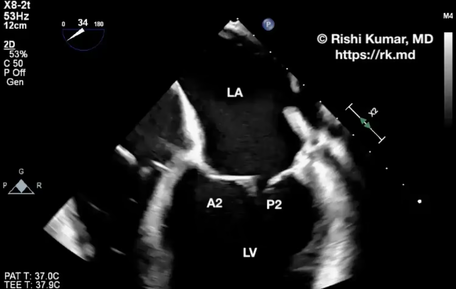
    Left ventricular (LV) dilation can stretch apart the mitral valve annulus causing the chordae tendinae to be pulled taut (particularly to the posterior leaflet). This restricts leaflet motion (Carpentier type III) over the cardiac cycle causing malcoaptation and associated mitral regurgitation (MR). Classically, with type III MR, the regurgitation jet…

    Read More

  • Surgical Septal Myectomy

    Surgical Septal Myectomy

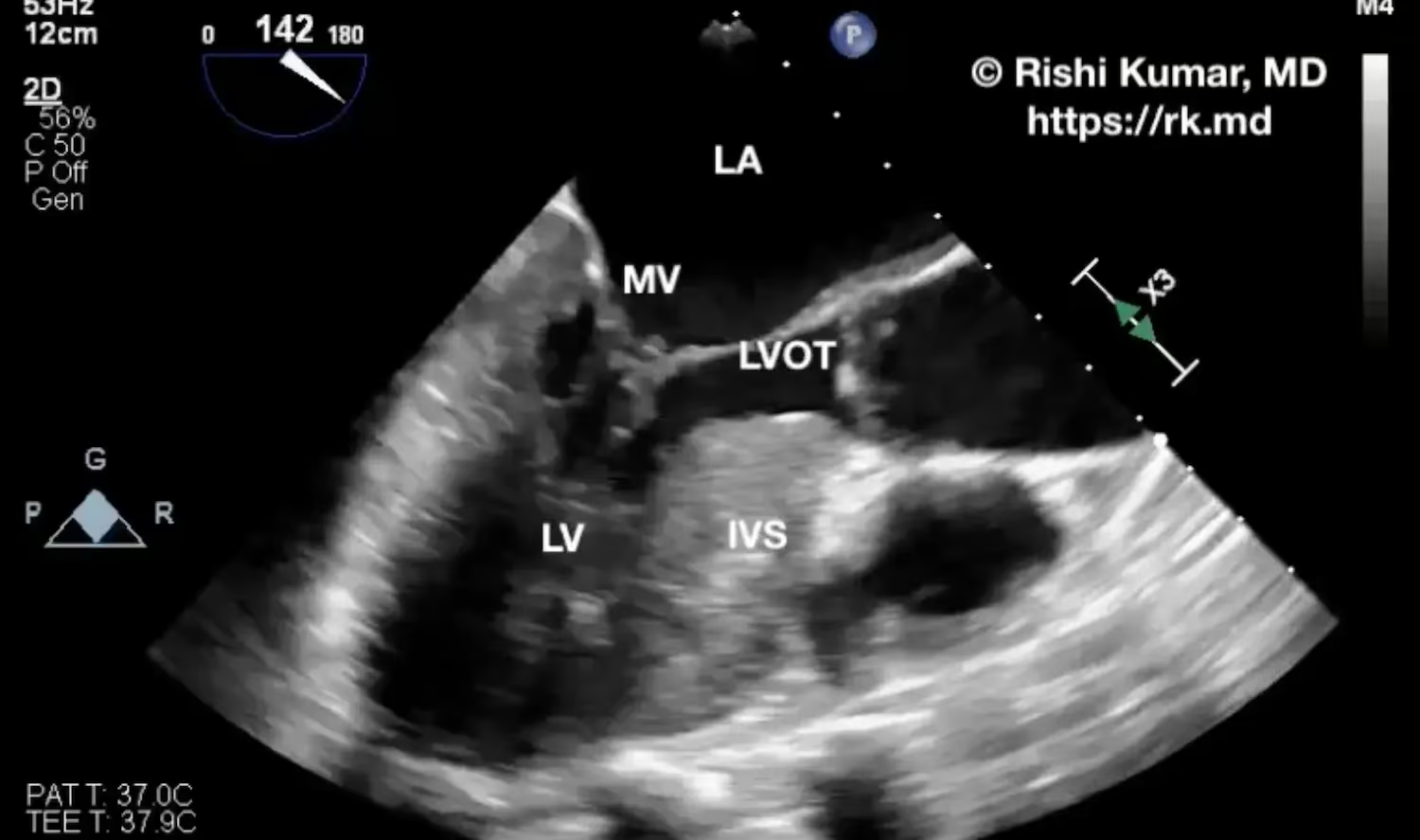
    Surgical septal myectomy is a procedure typically done for hypertrophic cardiomyopathy (HCM) where a portion of the hypertrophied interventricular septum (IVS) is resected. The basal portion of the IVS can lead to left ventricular outflow tract obstruction (LVOTO), limiting cardiac output and causing associated symptoms like dyspnea, lethargy, and organ…

    Read More