
A pneumothorax occurs when air fills the pleural space between the lungs and chest wall. This can cause partial or total lung collapse leading to difficulties with alveolar ventilation, hemodynamic lability, etc.
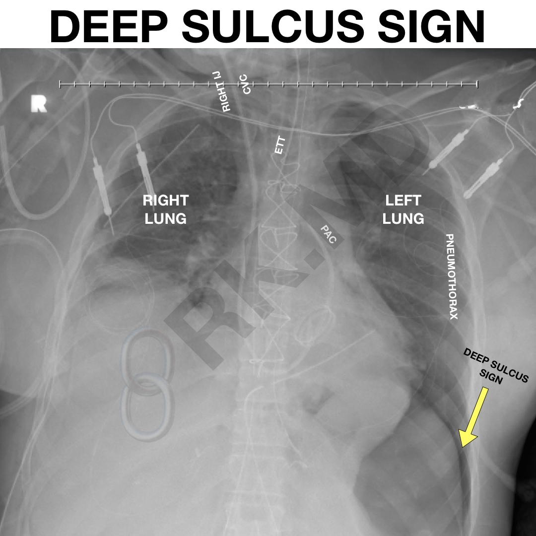
If a chest x-ray (CXR) is performed in the upright position, this air will rise and accumulate toward the apices of the lung fields. However, many ICU patients have portable A-P CXRs performed in the supine position. In this situation, free air shifts anteriorly and basally towards the non-dependent regions. Because air is distributed over a larger region in the pleural space, it sometimes makes it difficult to discern a pneumothorax on imaging.
One finding that suggests a pneumothorax is the deep sulcus sign. Notice how the left costophrenic region has an exaggerated extension downward. This is caused by air in the left lateral pleural space. The contour of the left hemidiaphragm becomes more prominent because air does not attenuate x-ray radiation as much as soft tissue. Indeed, this patient had a left-sided pneumothorax.
Drop me a comment below with questions!


