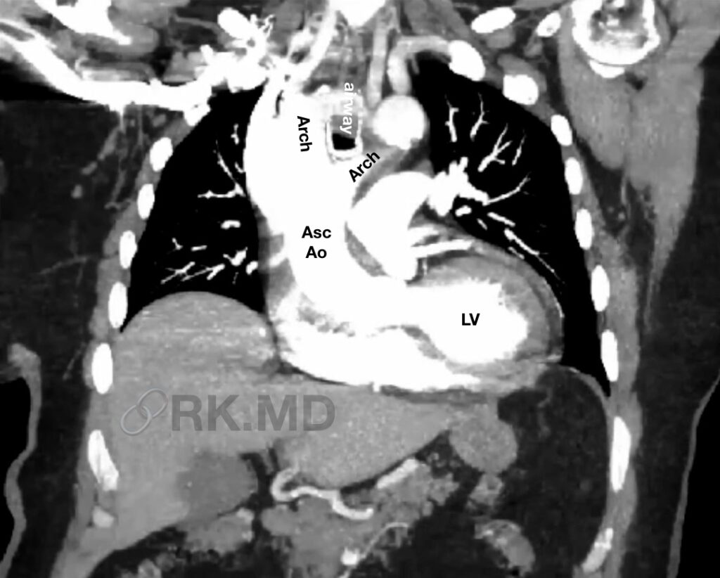
During embryogenesis, the aortic sac is connected to paired dorsal aortae via six symmetrical aortic (Ao) arches, which give rise to major vascular structures. For example, the right and left common carotid arteries originate from the right and left third arches, respectively. The proximal right subclavian artery is formed from the right fourth arch, and the left fourth arch forms the transverse aortic arch.


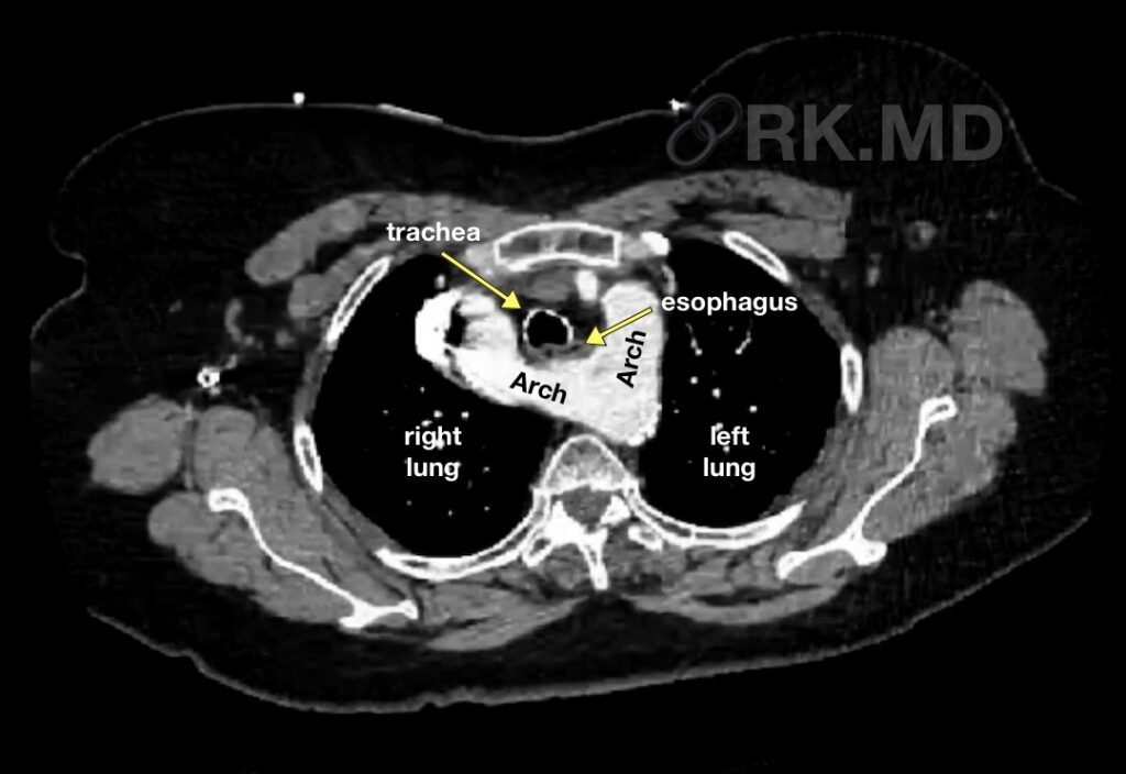
Double aortic arch (DAA) is a rare anomaly related to failed remodeling of the primitive fourth arch leading to two arches connecting the ascending aorta to the descending thoracic aorta. These two arches can form a complete vascular ring that entraps structures like the trachea and esophagus (as pictured on the CT), leading to symptoms of airway and/or esophageal compression like stridor, dysphagia, poor intake, etc.
Diagnosis of double aortic arch may be made through physical examination, medical history, and imaging tests such as echocardiogram, chest X-ray, and computed tomography (CT) scan. Treatment may involve surgical repair of the aorta to separate the two loops and relieve compression of the trachea and esophagus. The prognosis for individuals with double aortic arch is generally good with timely diagnosis and treatment.
Drop me a comment with questions!


