
The left atrial appendage (LAA) is a small pouch located within the pericardium close to the left ventricular free wall and in continuity with the upper body of the left atrium (LA). Due to its compliance and anatomic location, it serves as a decompression chamber when the LA pressure is elevated. In addition, atrial natriuretic peptide, a hormone that promotes sodium and water excretion, is also released by the LAA in response to hypertension and hypervolemia causing stretch.
Typically, blood moves in and out of the LAA during the cardiac cycle; however, in arrhythmias like atrial fibrillation (AFib), the LAA can become an area of stasis and clot formation. Because this is a left heart structure, the clot can embolize systemically leading to sequelae like strokes. Patients with non-valvular atrial fibrillation and a high CHA2DS2-VASc score warrant pharmacologic anticoagulation; however, a subset of these patients also have a high HAS-BLED score suggesting a major bleeding risk. Fortunately, options exist to eliminate flow to the LAA and allow patients to stop long-term anticoagulation.
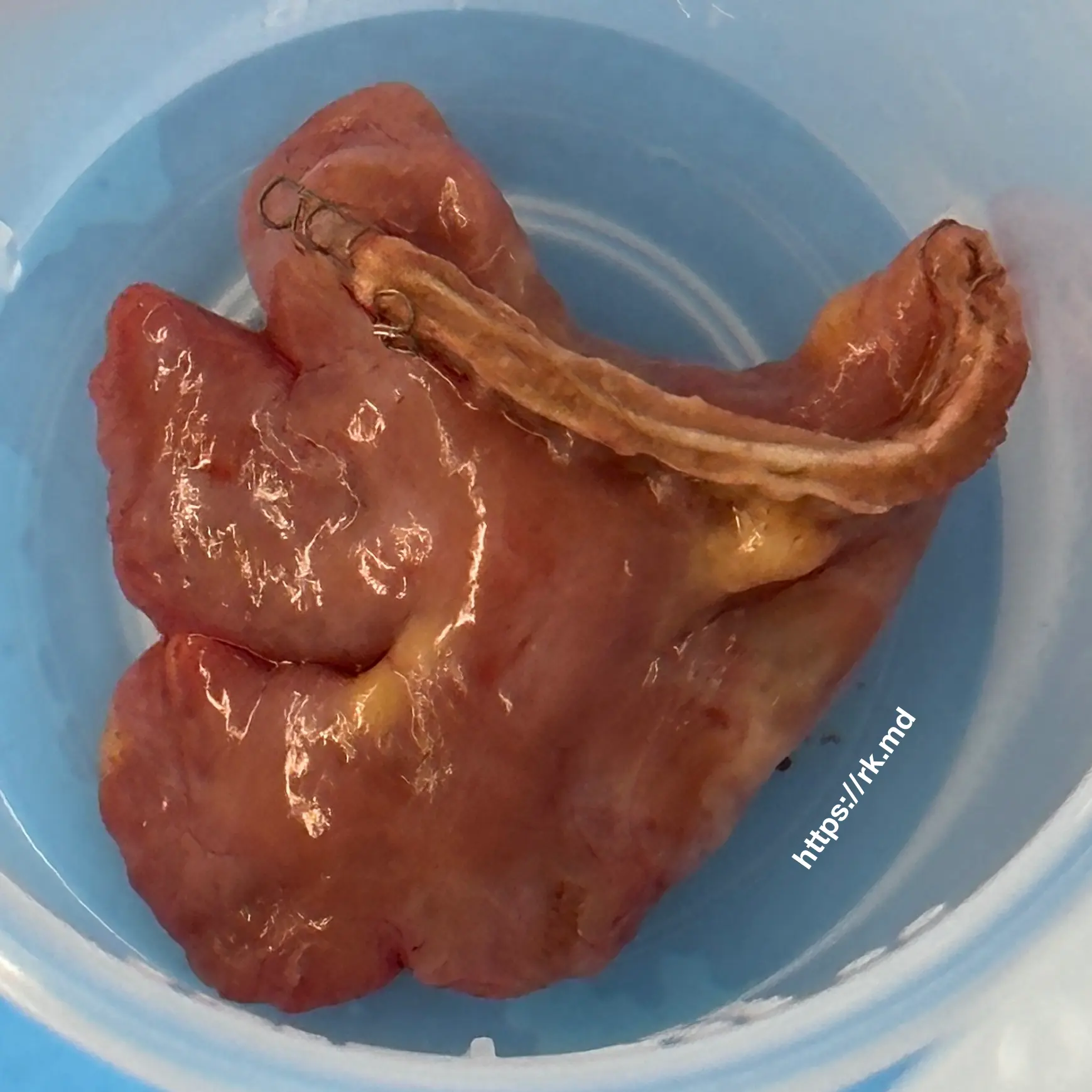
Cardiothoracic surgeons can perform various procedures, from basic LAA neck ligation and endocardial suturing (if the atrium is opened, for example, during mitral valve surgery) to clipping the LAA (e.g., AtriClip or LigaSure) versus excising it with a surgical stapler as pictured above. Cardiologists can achieve percutaneous occlusion of the LAA using devices like the Amulet or Watchman.
Have you seen any of these procedures at your institution? Drop me a comment below with questions and your experiences!


As an RN in a CTICU in a large NYC institution we received many patients who had LAA ligations. It was never the primary surgery but secondary to a valve replacement or some other cardiac case.
Absolutely! This is a fairly common practice when patients have an indication like atrial fibrillation.