• Left Ventricular Pseudoaneurysm

    Left Ventricular Pseudoaneurysm

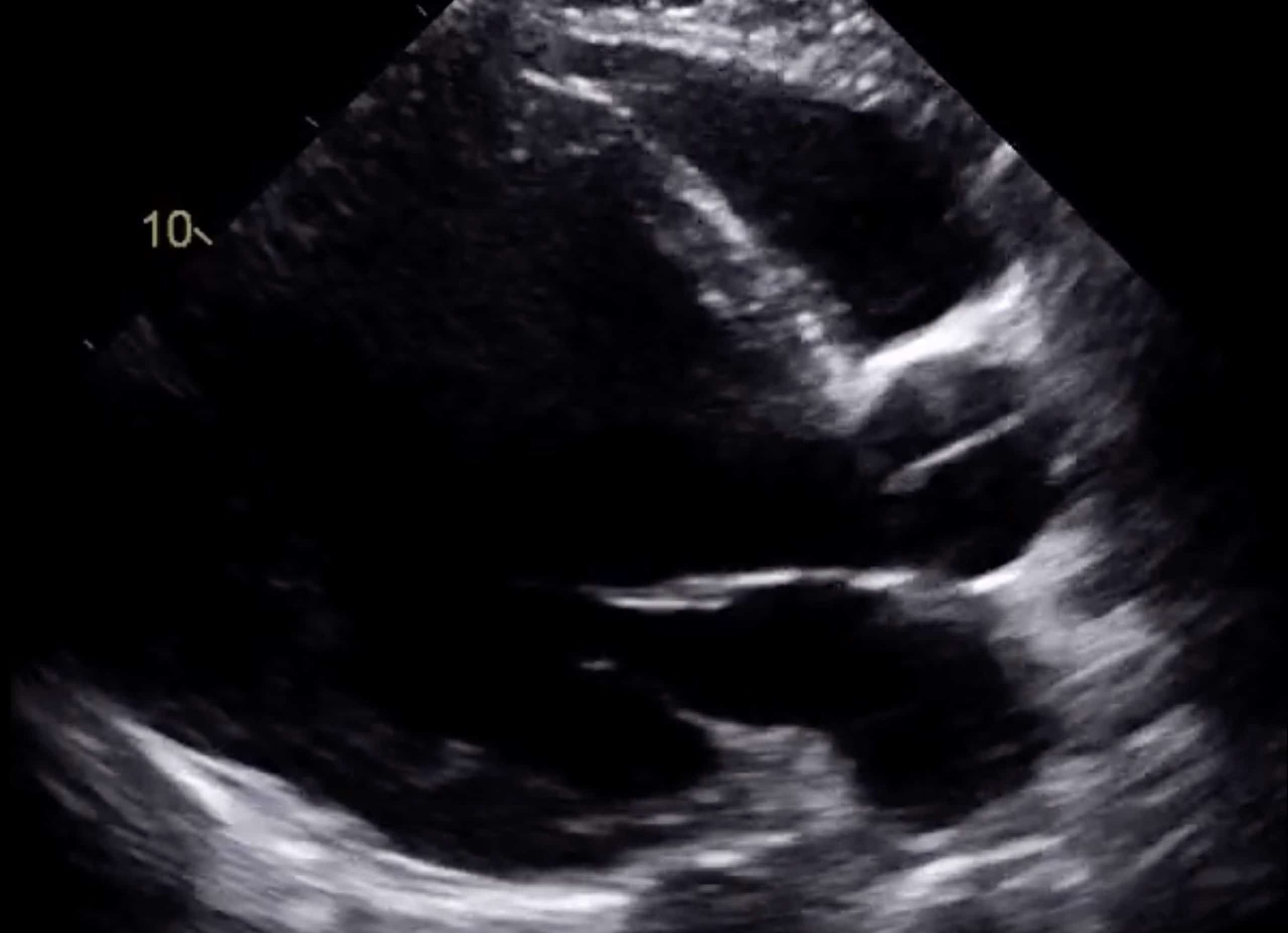
    A left ventricular (LV) pseudoaneurysm is an LV outpouching devoid of myocardium/endocardium contained by the pericardium and adhesions. As one can imagine, this pathology carries a high risk of rupture and mortality. LV pseudoaneurysms are most commonly linked to myocardial infarction (inferior > anterior) but can also follow open-heart surgery…

    Read More

  • Visipaque

    Visipaque

    Iodixanol (Visipaque) is an iodinated intravenous (IV) contrast agent which is iso-osmolar with plasma. Unlike the non-ionic monomer iohexol (Omnipaque), Visipaque is a non-ionic dimer; however, both agents are routinely utilized for angiography, venography, contrast-enhanced CT scans, and other diagnostic/interventional procedures. Tissue extravasation can result in localized necrosis, and certain…

    Read More

  • Ventricular Pacing Versus Normal Sinus Rhythm

    Ventricular Pacing Versus Normal Sinus Rhythm

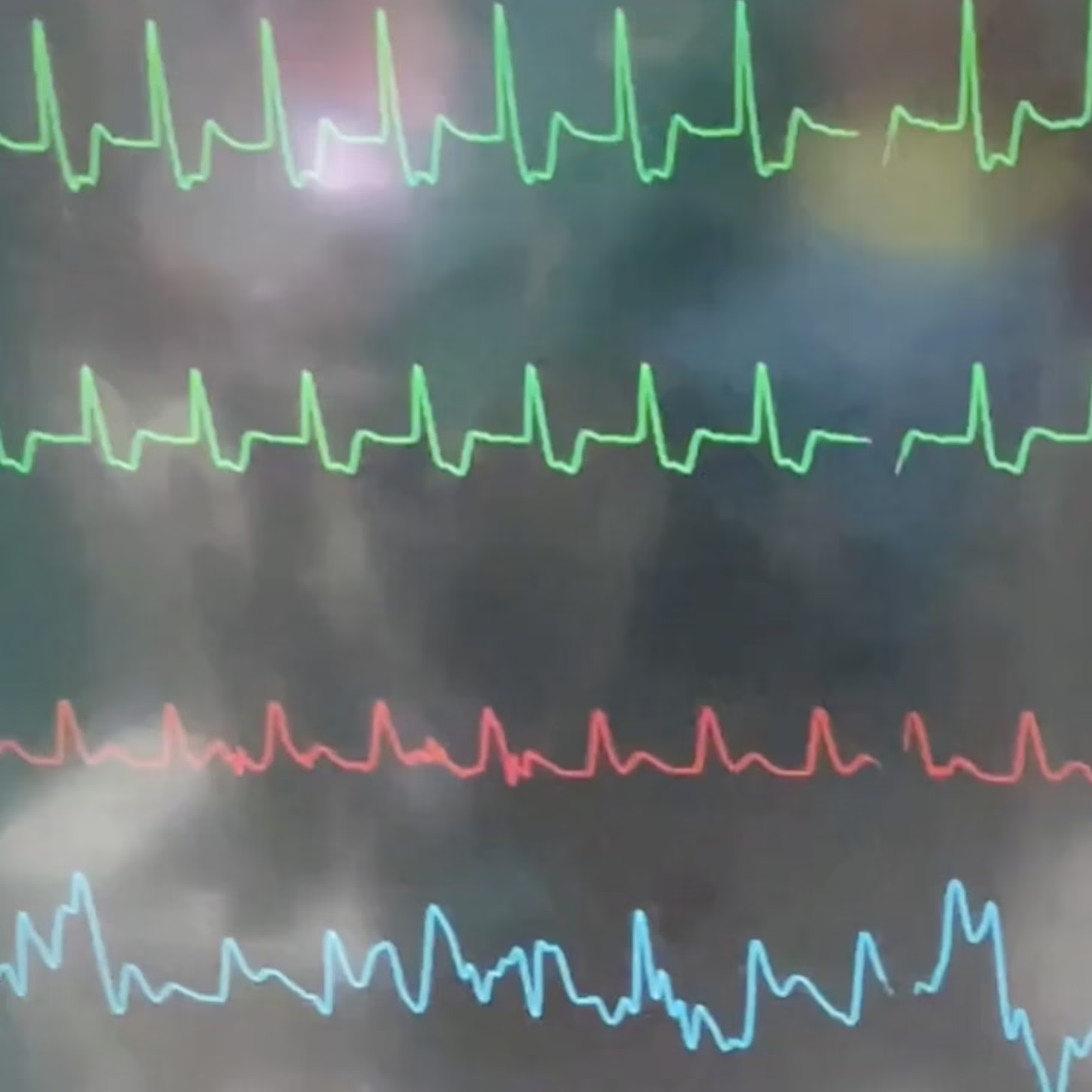
    As a cardiothoracic anesthesiologist and intensivist, many of my patients receive ventricular pacing at some point during their hospital admission. In the OR, many patients have epicardial right ventricular (RV) pacing implanted following cardiac surgery. In the ICU, I often encounter transvenous pacing (TVP) of the RV. Remember, normal cardiac…

    Read More

  • Anatomical Heart Emoji

    Anatomical Heart Emoji

    The “anatomical heart” was approved with Unicode 13.0 in 2020 and added to Emoji 13.0 the same year. Its appearance varies based on the platform (Apple, Google, Samsung, Facebook, etc), but below, I’ve labeled the iOS version with relevant anatomy… because I got bored one morning. 🤓

    Read More

  • S1-Q3-T3 ECG Pattern

    S1-Q3-T3 ECG Pattern

    S1-Q3-T3 refers to an ECG pattern first described in the 1930s suggestive of cor pulmonale in a case series of patients with pulmonary embolism (PE). This pattern has the following findings: It’s important to remember that this pattern is NOT pathognomonic for PE. In fact, S1-Q3-T3 should be LESS associated…

    Read More

  • Cardiac Action Potential

    Cardiac Action Potential

    In order for the heart muscle to actually contract, the physiological process of excitation-contraction coupling must occur via an action potential propagated through the myocardium. Normally, these muscle cells have a resting transmembrane potential (TMP) around -90 mV (more negative intracellularly than extracellularly). This gradient is created and changed by…

    Read More

  • The Pringles Mitral Valve

    The Pringles Mitral Valve

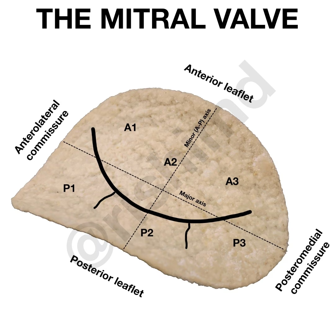
    The mitral valve’s complex geometry is usually described as a “saddle shape”, but I definitely prefer the comparison to Pringles chips! Here’s an illustration showing the relevant mitral anatomy. No Pringles were harmed in this production. :-)

    Read More

  • Don’t Fixate On Low Ejection Fraction!

    Don’t Fixate On Low Ejection Fraction!

    Early in my training as a medical student and intern, it seemed that all people ever cared about on an echo report was the “EF!” This refers to the left ventricular ejection fraction (LVEF) – the percentage of the left ventricle’s diastolic blood volume that is pumped out after a…

    Read More

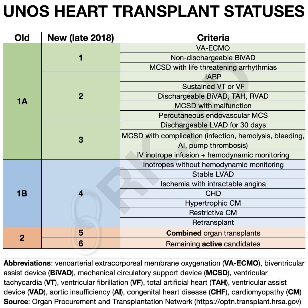
  • United Network for Organ Sharing (UNOS) 2018 Heart Transplant Listing Guidelines Revision
  • Alteplase

    Alteplase

    Alteplase (recombinant tissue plasminogen activator, tPA) is a potent intravenous fibrinolytic (“clot buster”) used for everything from acute ischemic strokes and clearing central line ports (“Cathflo”, a total life saver!) to hemodynamically unstable pulmonary embolism (PE) and even ST elevation myocardial infarctions (STEMIs – “heart attacks”).

    Read More