• Intracardiac Echocardiography (ICE)

    Intracardiac Echocardiography (ICE)

    Intracardiac echocardiography (ICE) allows cardiologists to visualize cardiac structures (aorta, valves, pulmonary veins, chambers, etc) from an ultrasound catheter usually threaded into the right atrium (RA) or right ventricle (RV) from the femoral vein. It aids in procedures ranging from percutaneous valve replacement and ablation to septal defect closures by…
    Read More

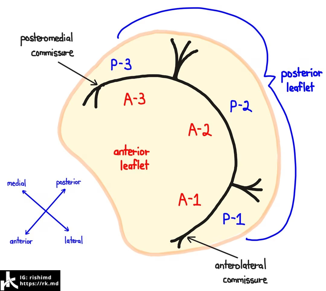
  • Mitral Valve Anatomy

    Mitral Valve Anatomy

    The mitral valve (MV) separates the left atrium (LA) and left ventricle (LV). It’s a bileaflet valve composed of an anterior and posterior leaflet, each with three “scallops” designated A1, A2, A3, P1, P2, and P3. The A3-P3 interface tapers into the posteromedial commissure whereas the A1-P1 interface becomes the…

    Read More

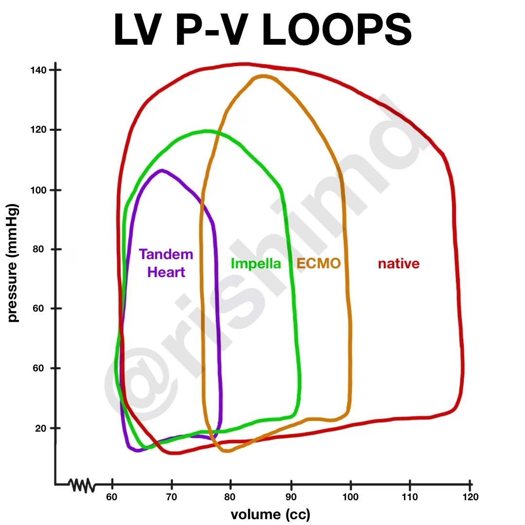
  • Left Ventricular Pressure-Volume Work Loop With Mechanical Circulatory Support
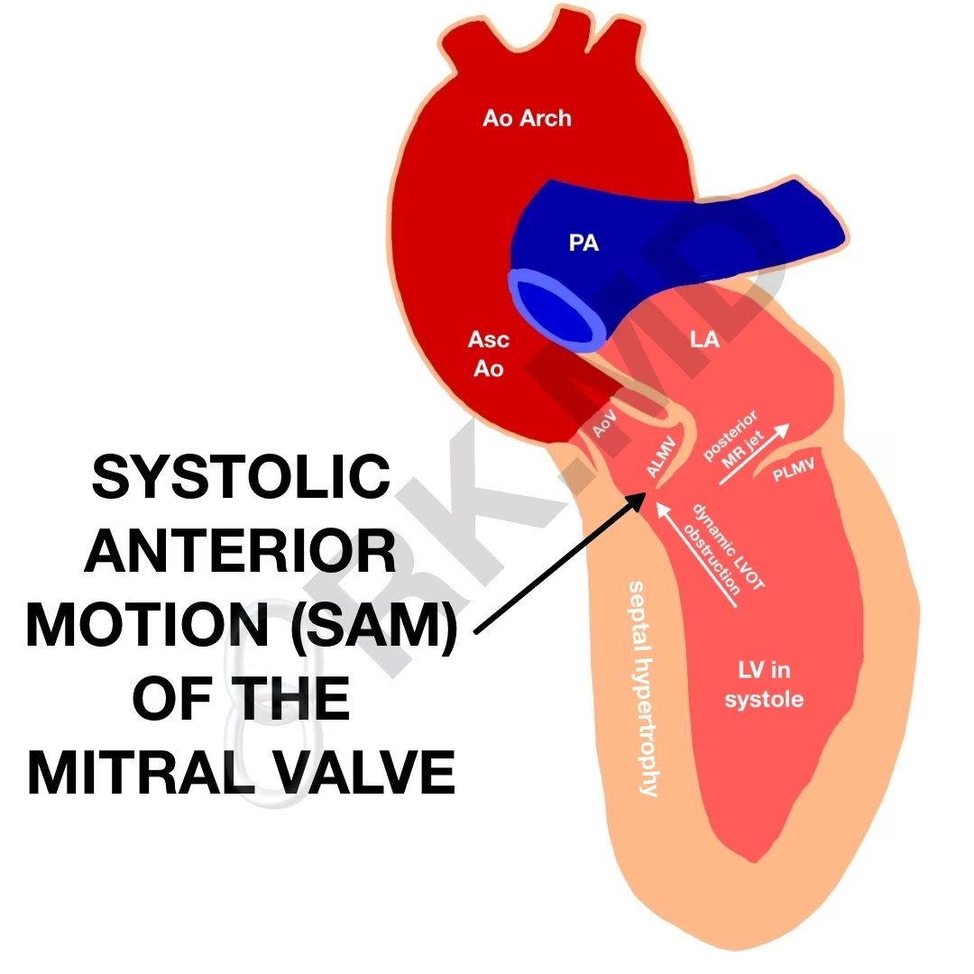
  • Systolic Anterior Motion (SAM) Of The Mitral Valve – Left Ventricular Outflow Tract (LVOT) Obstruction

    Systolic Anterior Motion (SAM) Of The Mitral Valve – Left Ventricular Outflow Tract (LVOT) Obstruction

    Left ventricular outflow tract (LVOT) obstruction is a potentially catastrophic etiology for hypotension due to several etiologies: idiopathic hypertrophic subaortic stenosis (IHSS), hypertrophic obstructive cardiomyopathy (HOCM), systolic anterior motion (SAM) of the mitral valve, and even mid-cavity obstructive hypertrophic cardiomyopathy. These obstructions can be dynamic in nature and consequently vary…

    Read More

  • What Is An Impella?

    What Is An Impella?

    Impella® is a percutaneous axial heart pump placed retrograde across the aortic valve into the left ventricle (LV) utilizing fluoroscopy +/- echocardiography. Its indications include high-risk percutaneous coronary intervention (PCI, especially cardiogenic shock or signs of ongoing ischemia), post-cardiotomy failure, and myocarditis. In addition, it is routinely used as a…

    Read More

  • What Is Cardiopulmonary Bypass (CPB)

    What Is Cardiopulmonary Bypass (CPB)

    This post outlines the steps in traditional, central cannulation for cardiopulmonary bypass (CPB). Depending on the nature of the surgery and the patient’s comorbidities, other sites are often cannulated (left heart bypass, femoral-femoral peripheral cannulation, etc.)

    Read More

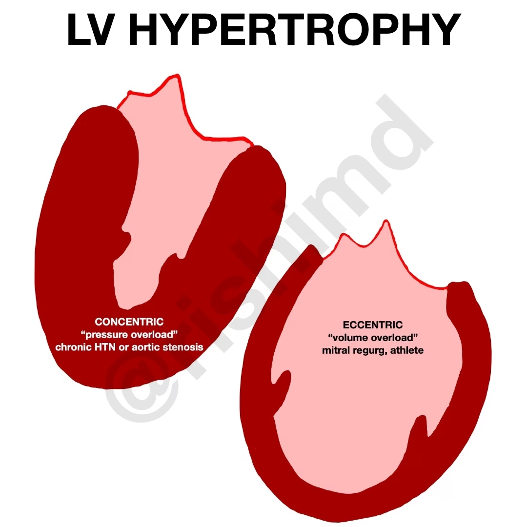
  • Eccentric Versus Concentric Hypertrophy
  • ECG Axis Deviation

    ECG Axis Deviation

    Normal cardiac conduction propagates downward and leftward from the SA node to the His-Purkinje system. Thus, lead I (points to the patient’s left) and lead aVF (points straight down) represent the component vectors in the x and y planes, respectively, for the cardiac conduction axis. This means that downward is + for lead aVF, and…

    Read More