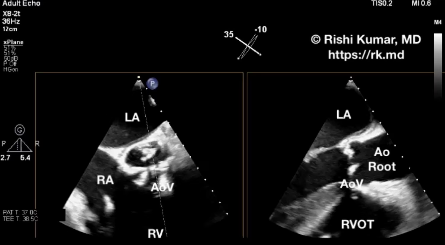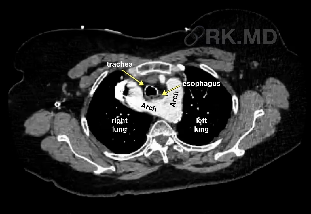
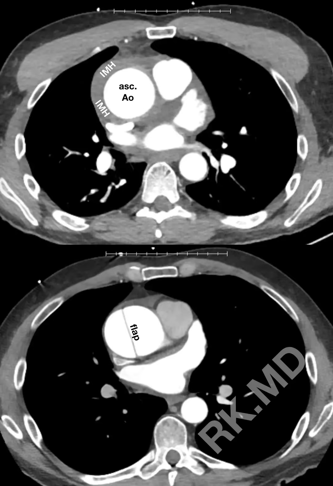
• Aortic Dissection Versus Intramural Hematoma (IMH)

    Aortic Dissection Versus Intramural Hematoma (IMH)

    Aortic dissection and intramural hematoma (IMH) are both acute aortic syndromes that can present similarly but have distinct pathophysiological differences and implications for management. For example, both can present with acute chest pain radiating to the back, often described as “tearing” or “ripping” in nature with or without hemodynamic instability.…
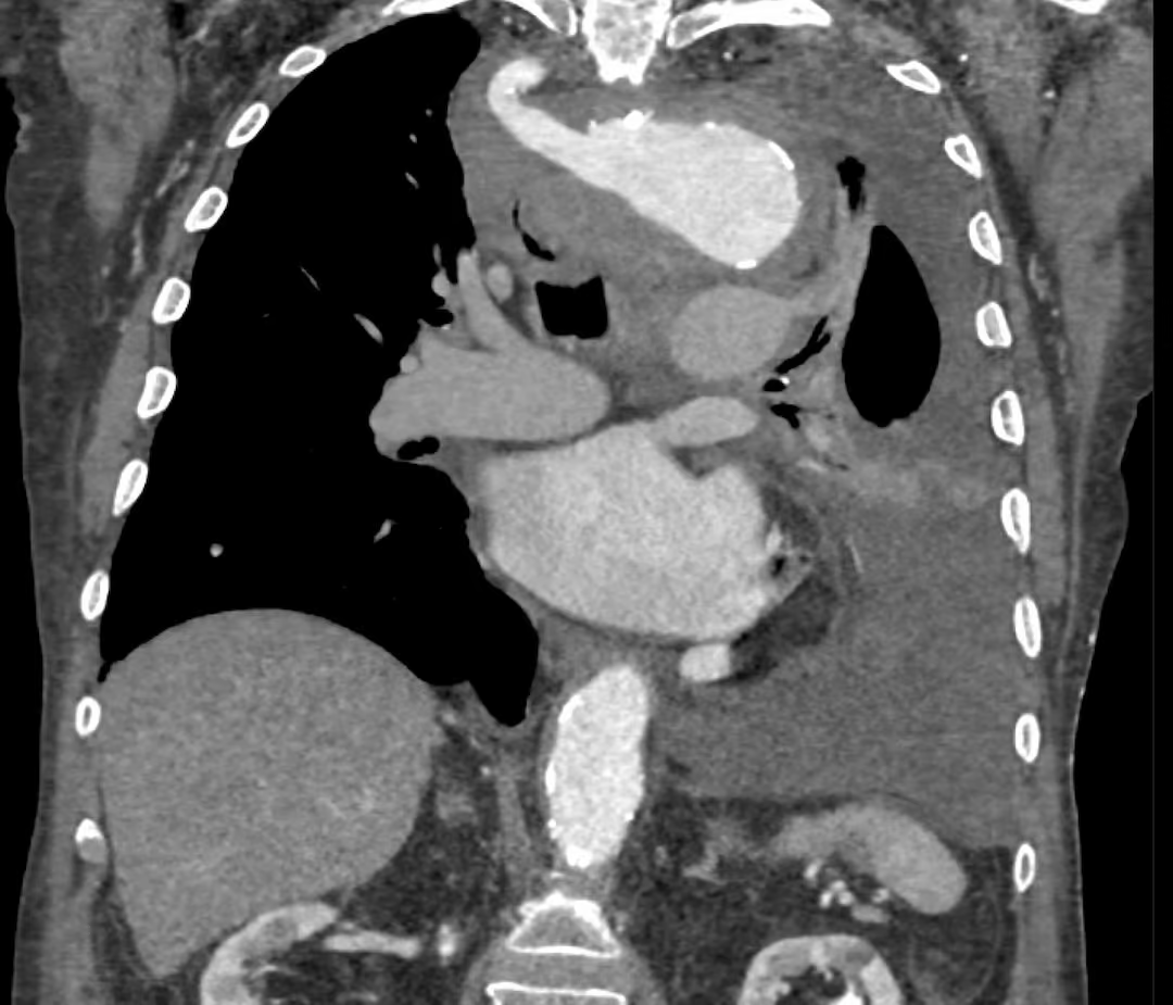
    Read More

  • Kommerell Diverticulum

    Kommerell Diverticulum

    Kommerell diverticulum (KD) is a rare vascular anomaly typically involving a right-sided aortic arch with an aberrant left subclavian artery. However, variations like a left-sided arch with an aberrant right subclavian artery exist. This condition results from the persistence of an embryonic vascular remnant – the dorsal segment of an…

    Read More

  • Marfan Syndrome

    Marfan Syndrome

    Marfan syndrome is a genetic condition primarily caused by mutations in the fibrillin-1 (FBN1) gene, which encodes a glycoprotein component of connective tissue. The manifestations of Marfan syndrome result from weakened connective tissues. These include everything from ocular (lens dislocation, retinal detachment, myopia) and cardiovascular (aortic aneurysm, aortic dissection, mitral…

    Read More

  • Aortic Dissection Flap Fenestrations

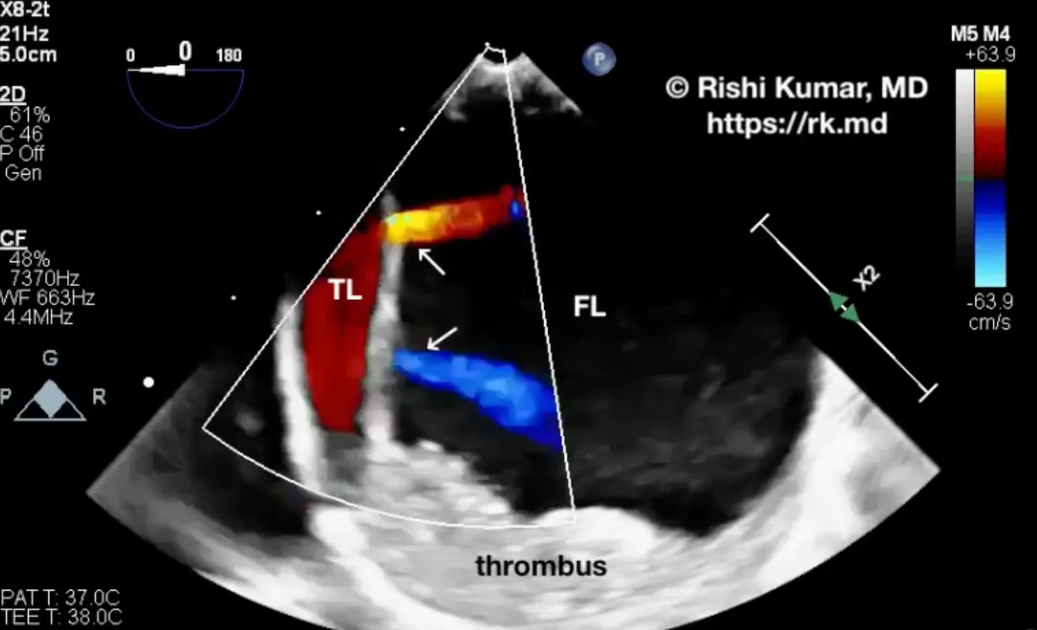
    Aortic Dissection Flap Fenestrations

    Aortic dissection is a life-threatening condition characterized by a tear in the innermost layer of the aorta (tunica intima), permitting blood to enter and separate the intima from the aorta’s middle layer (tunica media). This event creates a false lumen (FL) alongside the true lumen (TL) of the aorta. Typically,…

    Read More

  • Surgical Septal Myectomy

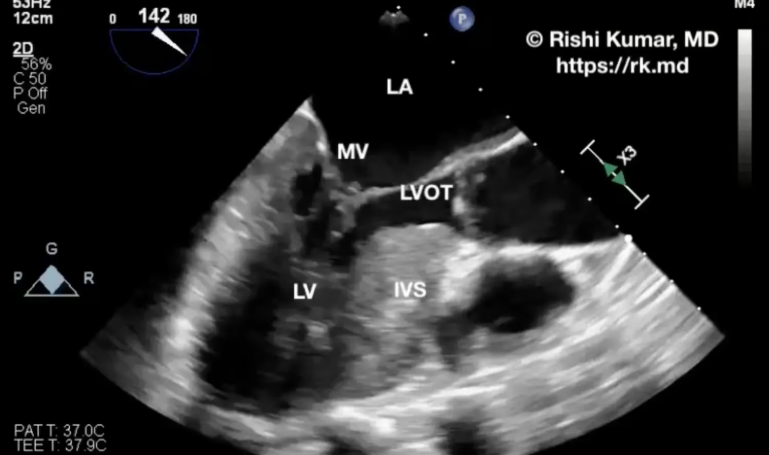
    Surgical Septal Myectomy

    Surgical septal myectomy is a procedure typically done for hypertrophic cardiomyopathy (HCM) where a portion of the hypertrophied interventricular septum (IVS) is resected. The basal portion of the IVS can lead to left ventricular outflow tract obstruction (LVOTO), limiting cardiac output and causing associated symptoms like dyspnea, lethargy, and organ…

    Read More

  • POCUS – Type B Aortic Dissection

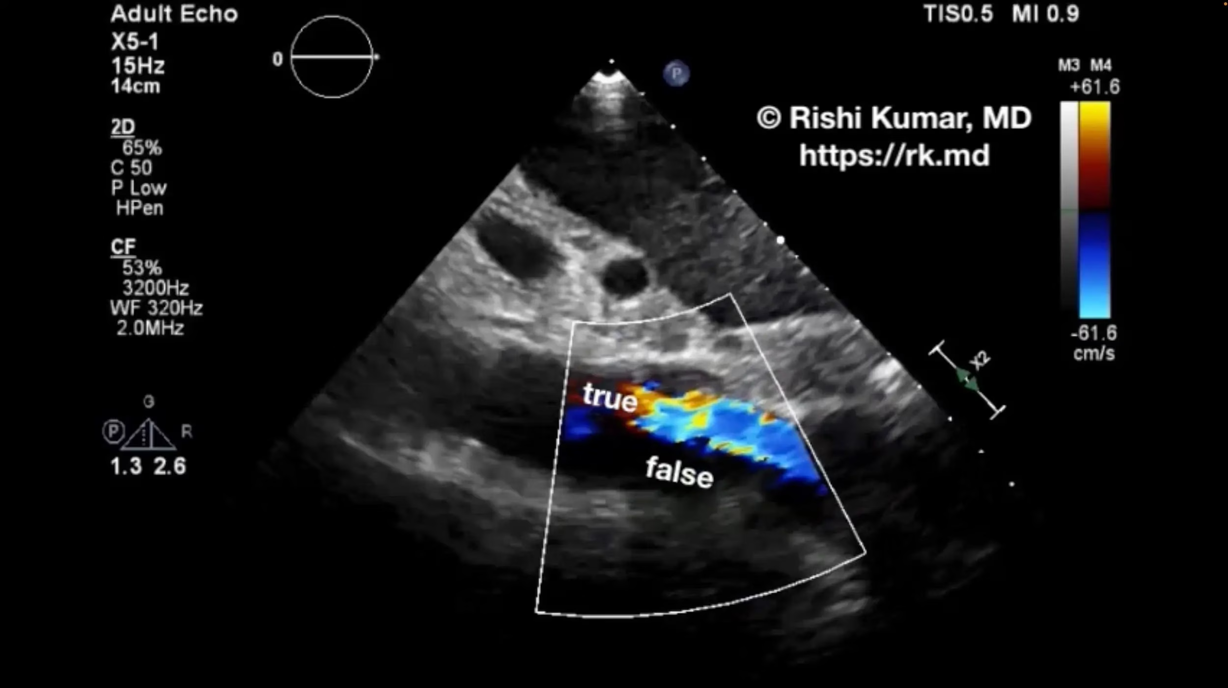
    POCUS – Type B Aortic Dissection

    Point-of-care ultrasound (POCUS) can be a quick and noninvasive modality to confirm a concerning pathology like type B aortic dissection. As a CVICU intensivist, I often receive transfers for “aortic dissection” from other hospitals based on suboptimal imaging. However, they’re sometimes found to be other acute aortic syndromes like intramural…

    Read More

  • Left Atrial Appendage Ligation

    Left Atrial Appendage Ligation

    The left atrial appendage (LAA) is a small pouch located within the pericardium close to the left ventricular free wall and in continuity with the upper body of the left atrium (LA). Due to its compliance and anatomic location, it serves as a decompression chamber when the LA pressure is…

    Read More

  • Bentall Procedure

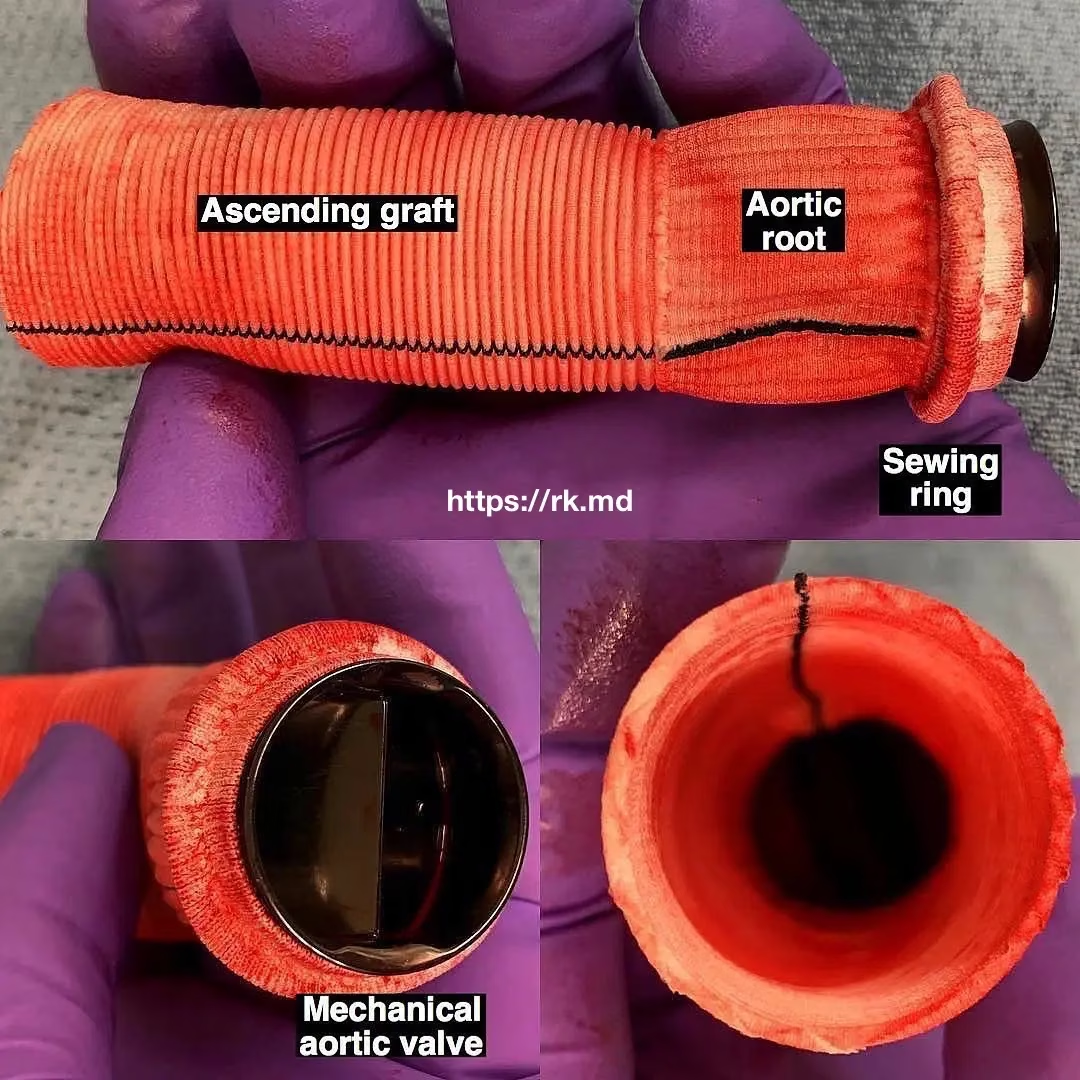
    Bentall Procedure

    Bentall procedure is a complex surgical procedure that involves the replacement of the aortic valve, ascending aorta, and aortic root with a prosthetic valve conduit. This procedure commonly treats patients with aortic aneurysms, aortic dissection, or aortic valve disease. The Bentall procedure was first introduced in 1968 by Dr. Hugh…

    Read More

  • Libman-Sacks Endocarditis

    Libman-Sacks Endocarditis

    Libman-Sacks endocarditis (LSE) is a nonbacterial endocarditis associated with lupus, malignancy, and antiphospholipid antibody syndrome characterized by endothelial injury and immune complex deposition typically on left-heart valves (mitral > aortic) causing inflammation and scarring. Unfortunately, symptoms are usually related to embolic sequelae such as stroke, abdominal pain related to gut…

    Read More

  • Double Aortic Arch

    Double Aortic Arch

    During embryogenesis, the aortic sac is connected to paired dorsal aortae via six symmetrical aortic (Ao) arches, which give rise to major vascular structures. For example, the right and left common carotid arteries originate from the right and left third arches, respectively. The proximal right subclavian artery is formed from…

    Read More