• Pulmonary Alveolar Proteinosis

    Pulmonary Alveolar Proteinosis

    Pulmonary alveolar proteinosis (PAP) is a diffuse lung disease caused by problems with: surfactant metabolism, alveolar macrophage function, granulocyte-macrophage colony stimulating factor, or an unknown etiology. These can lead to the deposition of surfactant and apoproteins within the alveoli. Symmetric alveolar opacities in a classic “bat-wing” distribution are characteristic chest…

    Read More

  • Left Ventricular Pseudoaneurysm

    Left Ventricular Pseudoaneurysm

    A left ventricular (LV) pseudoaneurysm is an LV outpouching devoid of myocardium/endocardium contained by the pericardium and adhesions. As one can imagine, this pathology carries a high risk of rupture and mortality. LV pseudoaneurysms are most commonly linked to myocardial infarction (inferior > anterior) but can also follow open-heart surgery…

    Read More

  • Visipaque

    Visipaque

    Iodixanol (Visipaque) is an iodinated intravenous (IV) contrast agent which is iso-osmolar with plasma. Unlike the non-ionic monomer iohexol (Omnipaque), Visipaque is a non-ionic dimer; however, both agents are routinely utilized for angiography, venography, contrast-enhanced CT scans, and other diagnostic/interventional procedures. Tissue extravasation can result in localized necrosis, and certain…

    Read More

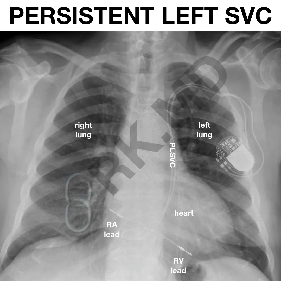
  • Persistent Left Superior Vena Cava (SVC)

    Persistent Left Superior Vena Cava (SVC)

    Persistent left superior vena cava (PLSVC) is a rare (~0.5% of the general population) venous anomaly when the left superior cardinal vein fails to degenerate. This can make entering the right heart from the left upper body (e.g., pacing leads, PA catheters, etc.) more challenging as the PLSVC drains directly…

    Read More

  • Cerebral Perfusion Scans And Brain Death

    Cerebral Perfusion Scans And Brain Death

    Cerebral perfusion scans are nuclear medicine studies utilizing agents like DTPA or NEUROLITE to confirm a clinical diagnosis of brain death. NEUROLITE is a radioactive diagnostic agent (technetium Tc99m bicisate) which passively diffuses across the intact blood-brain barrier and remains stable in the brain for ~6 hours. As such, this…

    Read More

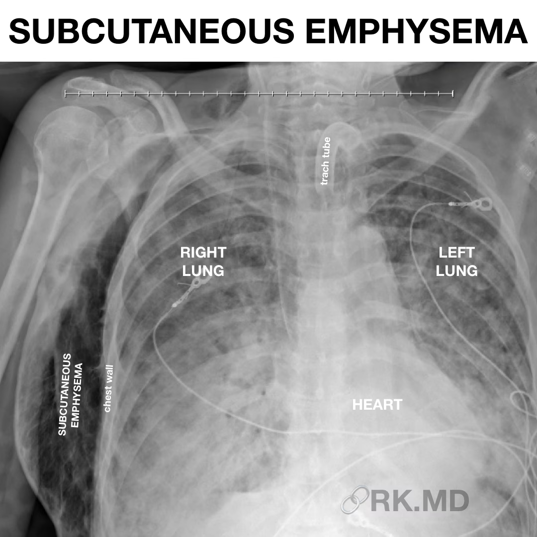
  • Subcutaneous Emphysema

    Subcutaneous Emphysema

    Subcutaneous emphysema occurs when air gets trapped within the subcutaneous regions of the face, neck, chest wall, abdomen, and even down to the thighs/legs. The characteristic physical exam finding of crepitus (“snap-crackle-pop”) is caused by air traversing tissue planes during palpation.

    Read More

  • Prone Chest X-Rays

    Prone Chest X-Rays

    In severe COVID-19 pneumonia with refractory hypoxemia, patients are often proned (put on their abdomen), deeply sedated, and paralyzed to improve synchrony with the ventilator. The goal with proning is to create a more homogenous alveolar recruitment thereby decreasing shunting (parts of the lung where there is pulmonary blood flow…

    Read More

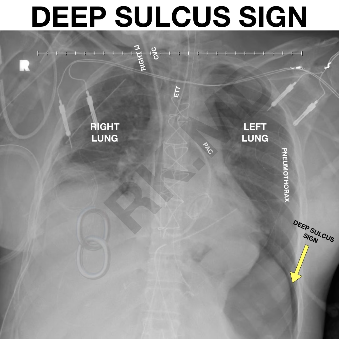
  • Deep Sulcus Sign

    Deep Sulcus Sign

    A pneumothorax occurs when air fills the pleural space between the lungs and chest wall. This can cause partial or total lung collapse leading to difficulties with alveolar ventilation, hemodynamic lability, etc.

    Read More

  • Tension Pneumothorax

    Tension Pneumothorax

    Here’s an example of a large, right-sided tension pneumothorax (PTX). Disruption in the lung leads to a one-way valve where air fills the pleural space and expands the PTX during inspiration but cannot escape during expiration.

    Read More

  • Air Bronchograms

    Air Bronchograms

    In this portable x-ray of a patient with COVID-19 pneumonia, one can clearly see the right and left mainstem bronchi as well as more distal segments (arrows indicate several “air bronchograms”). This occurs because the air-filled, patent bronchi don’t attenuate x-ray radiation and appear dark compared to the surrounding airspace…

    Read More