
Persistent left superior vena cava (PLSVC) is a rare (~0.5% of the general population) venous anomaly when the left superior cardinal vein fails to degenerate. This can make entering the right heart from the left upper body (e.g., pacing leads, PA catheters, etc.) more challenging as the PLSVC drains directly into the coronary sinus (CS) rather than the right atrium (RA).
During cardiac surgery, if I see an enlarged coronary sinus on transesophageal echocardiography (especially if there is no other reason for the RA pressure to be elevated), I have to consider the presence of a persistent left SVC. Agitated saline contrast injected in a left arm vein will lead to contrast in the CS before the RA. If this is the case, retrograde cardioplegia (given in the CS) won’t be a great option to arrest the heart as the cardioplegia solution travels systemically via the PLSVC rather than staying within the coronary venous system.
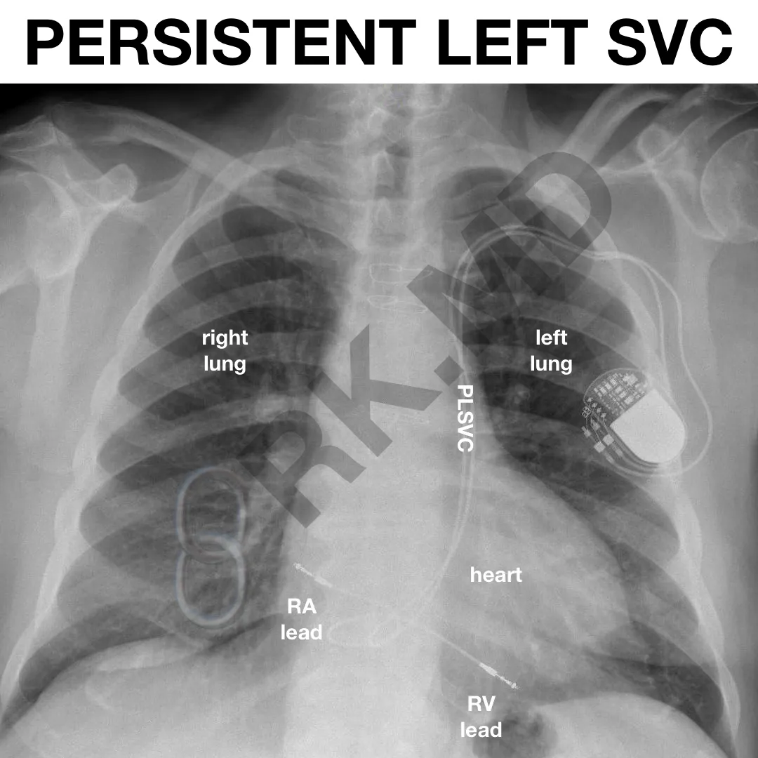
This chest x-ray shows a dual-chamber pacemaker’s leads traveling through a PLSVC to the heart.
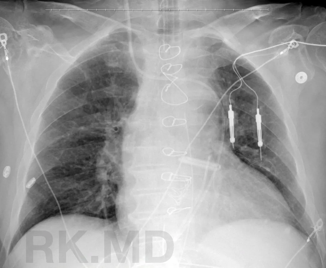
Here’s another example where a Swan-Ganz catheter placed from the right internal jugular vein is coursing through a persistent left SVC → coronary sinus → right atrium → right ventricle → pulmonary artery.
Finally, here’s an example of a TEE video clip (provided by my colleague, Dr. Sanjana Malviya) which illustrates a dilated coronary sinus (CS) in the midesophageal 4-chamber view just before entering the transgastric reference point. This raises suspicion for a PLSVC.
Have you seen PLSVC? Drop me a comment with your experiences and questions!


