• Boston Perioperative Echocardiography Presentation

    Boston Perioperative Echocardiography Presentation

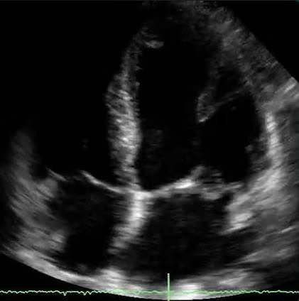
    To kick off the 16th year of Boston’s perioperative echocardiography presentations, I had the privilege of representing my program yesterday evening at Tufts. I discussed Shone’s Syndrome and relevant TTE/TEE imaging from a case. Having the support of my faculty and colleagues was incredible – it made me even more…

    Read More

  • The Continuity Equation

    The Continuity Equation

    The continuity equation illustrates the conservation of energy. In the case of blood flow within the (normal) heart, volumetric flow (cardiac output) at serial sites should be equal. The image shows flow velocities across the left ventricular outflow tract (LVOT) and through the aortic valve. Because these structures are in…

    Read More

  • The Doppler Effect

    The Doppler Effect

    The Doppler effect refers to a shift in frequency perceived by an object in motion relative to the source of a wave. When applying this to ultrasonography, the transducer is a stationary source of ultrasound waves of a known frequency (depending on the mode) and velocity (1540 meters/second) which reflect…

    Read More

  • Epiaortic Ultrasound (EAS)

    Epiaortic Ultrasound (EAS)

    Today’s didactic lecture on epiaortic ultrasonography was given by one of my wonderful cardiothoracic anesthesiology attendings – Dr. Jeff Swanson. As part of traditional cardiac surgeries, the aorta can be extensively manipulated – especially when performing arterial cannulation and applying the aortic cross-clamp. This can dislodge plaques systemically leading to…

    Read More

  • Left Atrial Spontaneous Echo Contrast (SEC)

    Left Atrial Spontaneous Echo Contrast (SEC)

    Left atrial spontaneous echo contrast (SEC) is an echogenic, smoke-like swirling of aggregated red blood cells suggestive of stasis. This sign confers an increased risk of thrombosis in the left atrium and/or left atrial appendage. We most commonly see SEC on transesophageal echocardiography (TEE) in patients with atrial arrhythmias like…

    Read More

  • Intracardiac Echocardiography (ICE)

    Intracardiac Echocardiography (ICE)

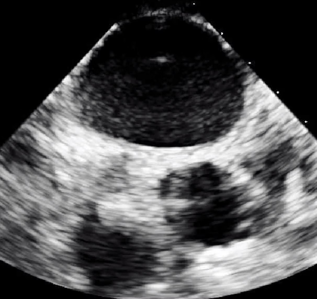
    Intracardiac echocardiography (ICE) allows cardiologists to visualize cardiac structures (aorta, valves, pulmonary veins, chambers, etc) from an ultrasound catheter usually threaded into the right atrium (RA) or right ventricle (RV) from the femoral vein. It aids in procedures ranging from percutaneous valve replacement and ablation to septal defect closures by…

    Read More

  • Mitral Valve Anatomy

    Mitral Valve Anatomy

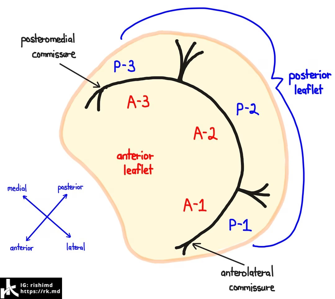
    The mitral valve (MV) separates the left atrium (LA) and left ventricle (LV). It’s a bileaflet valve composed of an anterior and posterior leaflet, each with three “scallops” designated A1, A2, A3, P1, P2, and P3. The A3-P3 interface tapers into the posteromedial commissure whereas the A1-P1 interface becomes the…

    Read More

  • Ultrasound Pleural Effusion And Spine Sign

    Ultrasound Pleural Effusion And Spine Sign

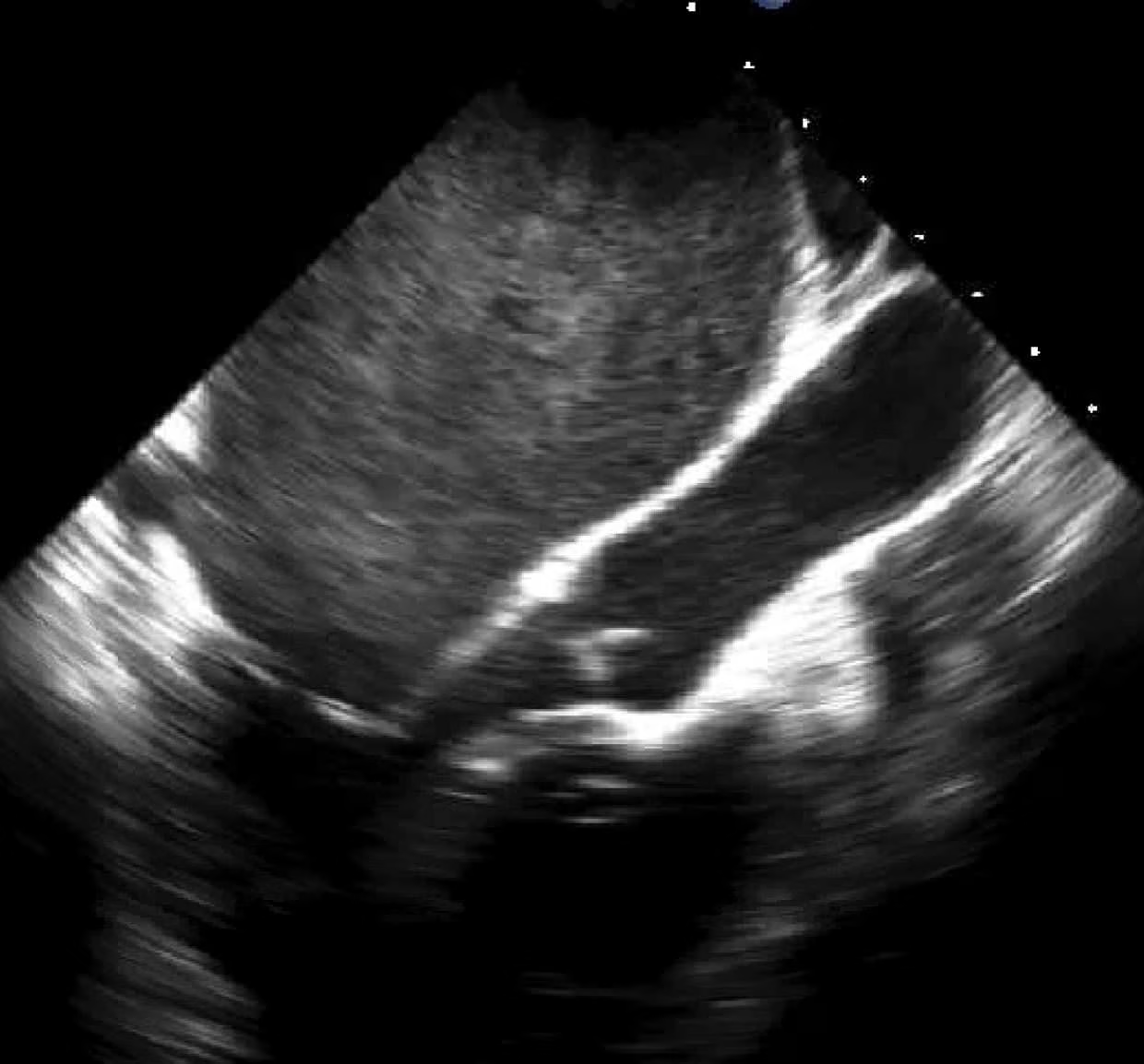
    In this ultrasound still image, I have a phase array probe positioned on the left side of the body around the 8th rib. A left-sided pleural effusion is noted. It’s important to appreciate this view to look for free fluid in the subphrenic and splenorenal spaces. The green arrow denotes…

    Read More

  • Systolic Anterior Motion (SAM) Of The Mitral Valve – Left Ventricular Outflow Tract (LVOT) Obstruction

    Systolic Anterior Motion (SAM) Of The Mitral Valve – Left Ventricular Outflow Tract (LVOT) Obstruction

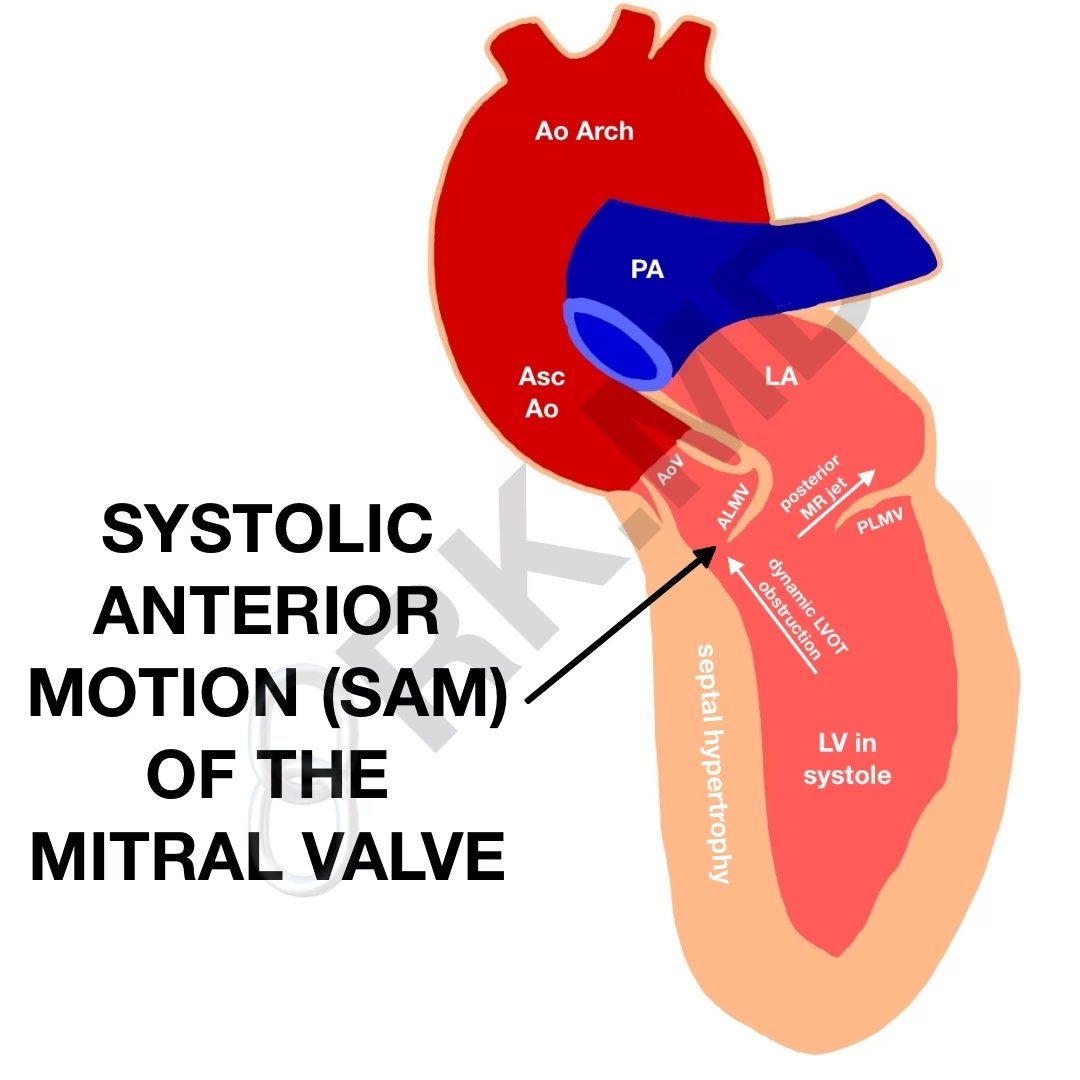
    Left ventricular outflow tract (LVOT) obstruction is a potentially catastrophic etiology for hypotension due to several etiologies: idiopathic hypertrophic subaortic stenosis (IHSS), hypertrophic obstructive cardiomyopathy (HOCM), systolic anterior motion (SAM) of the mitral valve, and even mid-cavity obstructive hypertrophic cardiomyopathy. These obstructions can be dynamic in nature and consequently vary…

    Read More

  • Cardiac Transthoracic Echocardiography (TTE) – Summary And Labeled Views

    Cardiac Transthoracic Echocardiography (TTE) – Summary And Labeled Views

    Here’s a quick primer of cardiac transthoracic echocardiography (TTE) including actual images I’ve taken as a critical care fellow with corresponding labeled diagrams. I challenge you all to actually review your patients’ echo images instead of reading the final impression. You don’t need to know the nitpicky numbers, gradient assessments,…

    Read More