
Right-sided aortic arch (RAA) is a rare (~0.1% of the population) anatomical variation of the aorta’s position and branching pattern. It is associated with congenital heart defects (truncus arteriosus, Tetralogy of Fallot) and chromosomal abnormalities (DiGeorge syndrome, Down syndrome). There are several types of right-sided aortic arch, each characterized by specific features and associated anomalies:
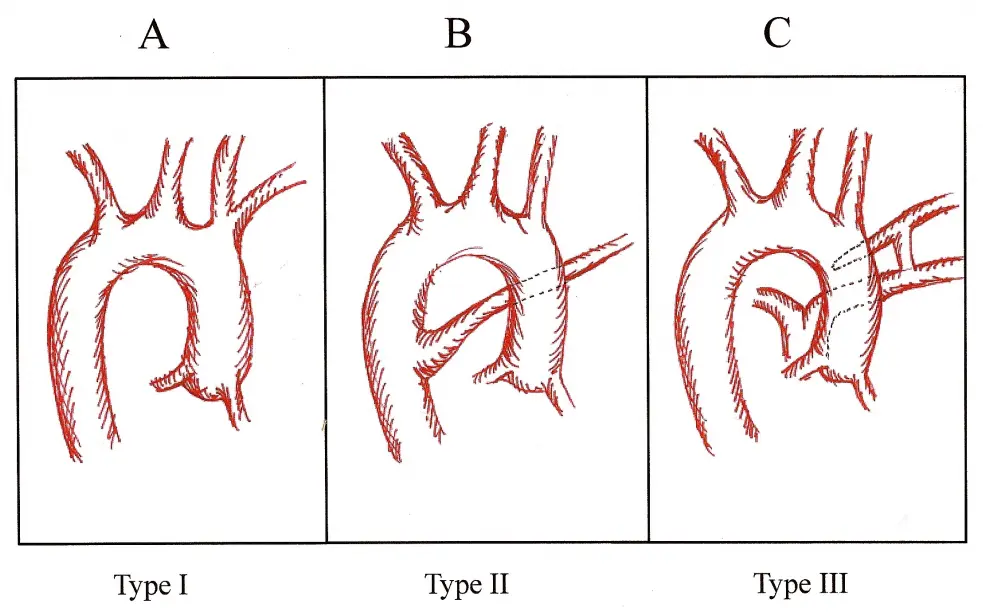
- Type I: most common RAA configuration in which the arch vessels mirror the usual anatomy resulting in the following branches (proximal to distal): left innominate artery (IA), right common carotid (RCC) artery, and right subclavian artery (RSA). The descending thoracic aorta (DTA) courses along the right side of the spine.
- Type II: second most common configuration – left carotid artery, right carotid artery, right subclavian artery, and an aberrant left subclavian artery (ALSA). The RAA + ALSA can create a vascular ring and compression of the trachea and esophagus.
- Type III: rarest configuration – like type II, but instead of an ALSA, the left subclavian artery is connected to the pulmonary artery by a left ductus arteriosus.
RSA might not always present with evident symptoms; however, its associations and implications warrant work-up. Additionally, the potential for vascular ring anomalies can lead to respiratory and feeding difficulties, especially in infants.
Diagnosis is centered around imaging techniques like echocardiography, CT, and MRI. Once identified, the management approach varies depending on associated anomalies and symptoms. Surgical intervention may be required to address vascular ring anomalies or other cardiac defects.


