• Doppler Shift – The Angle Matters!

    Doppler Shift – The Angle Matters!

    The Doppler shift equation describes the observed change in frequency or wavelength of a wave (such as sound, light, or radio waves) when the source of the wave and the observer are in relative motion. This phenomenon is known as the Doppler effect and is incredibly important to understand when…
    Read More

  • Tetralogy Of Fallot (TOF)

    Tetralogy Of Fallot (TOF)

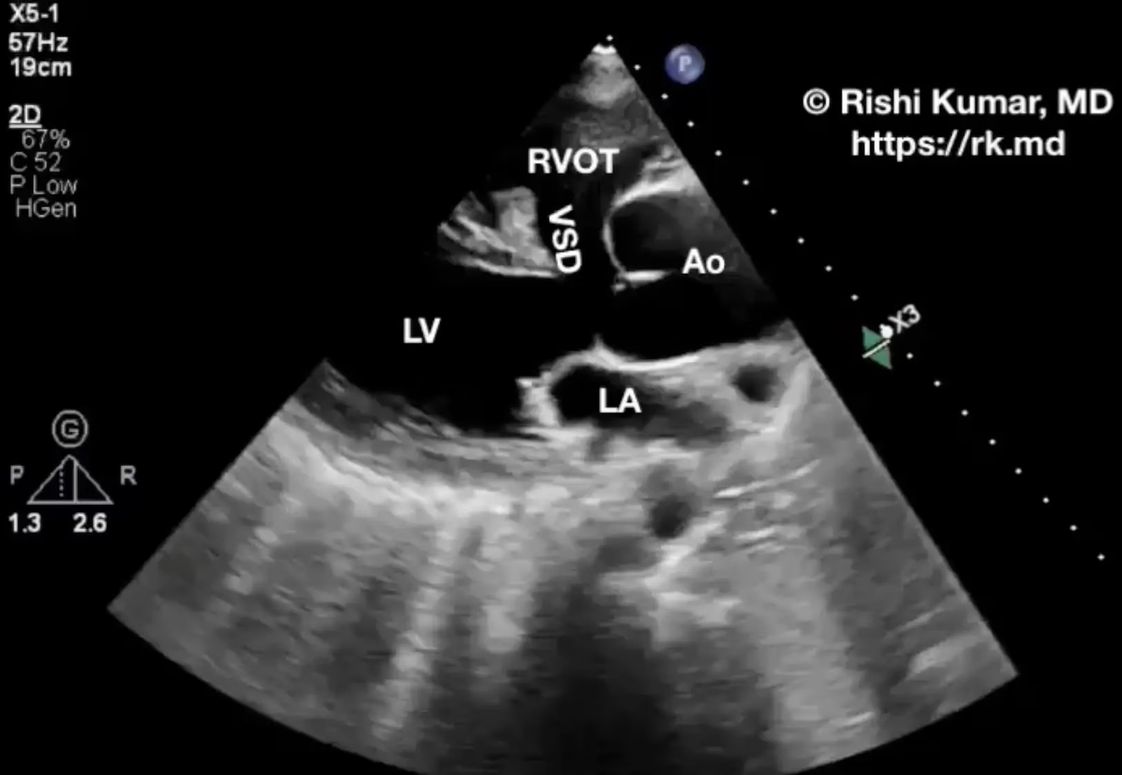
    Tetralogy Of Fallot (TOF), named for French physician Arthur Fallot, is the most common cyanotic congenital heart defect and comprised of four cardiovascular anomalies: RVOT obstruction reduces blood flow to the lungs resulting in chronic hypoxemia and right-to-left shunting through the VSD. It’s common for these patients to have hemoglobin…

    Read More

  • Right-Sided Aortic Arch (RAA)

    Right-Sided Aortic Arch (RAA)

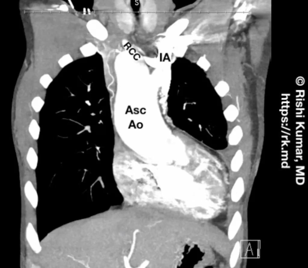
    Right-sided aortic arch (RAA) is a rare (~0.1% of the population) anatomical variation of the aorta’s position and branching pattern. It is associated with congenital heart defects (truncus arteriosus, Tetralogy of Fallot) and chromosomal abnormalities (DiGeorge syndrome, Down syndrome). There are several types of right-sided aortic arch, each characterized by…

    Read More

  • The Apnea Test

    The Apnea Test

    Brain death is a clinical diagnosis supported by imaging but confirmed by the bedside exam to assess for the irreversible loss of cortical and brainstem functions. The apnea test (AT) is a common method used to evaluate the definitive absence of a brainstem-mediated respiratory drive. To ensure patient safety and…

    Read More

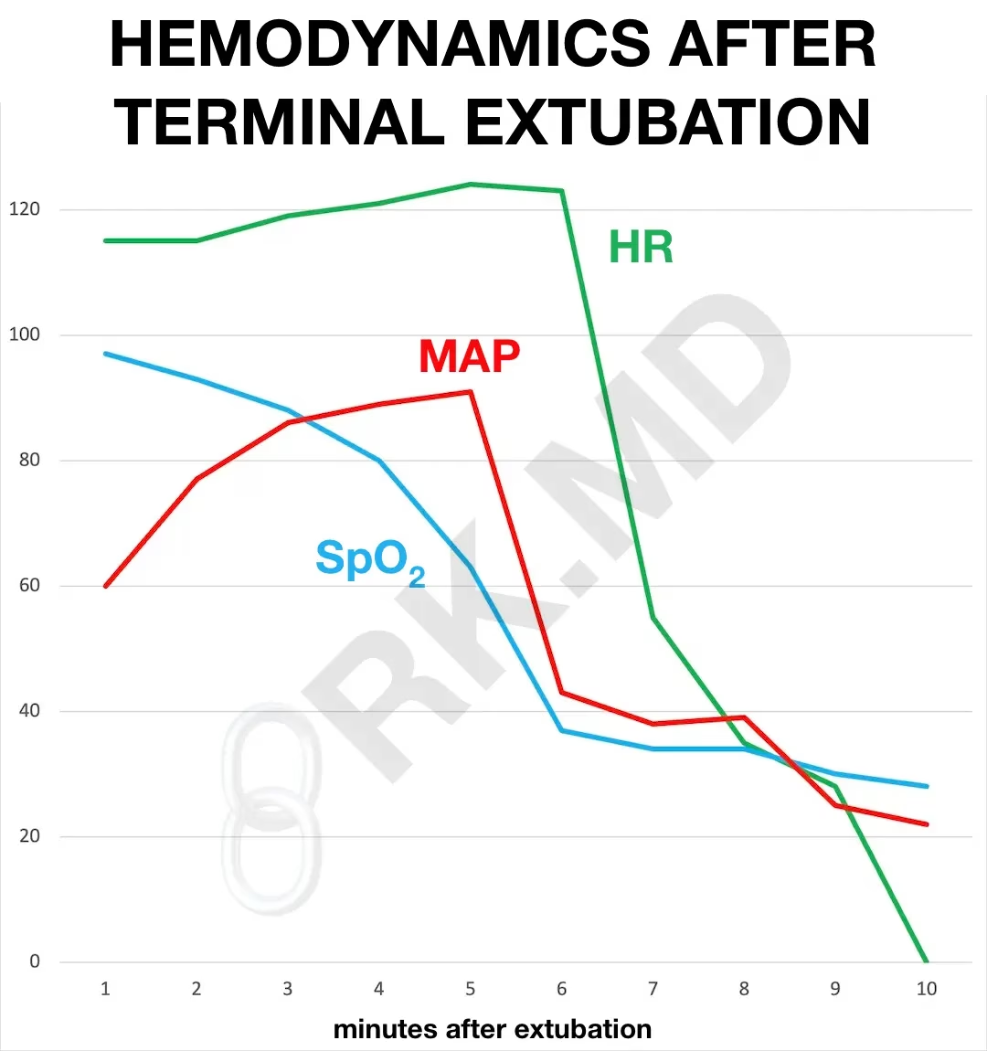
  • Hemodynamic Changes After Terminal Extubation

    Hemodynamic Changes After Terminal Extubation

    Removing an endotracheal tube and ventilator support from a patient nearing the end of life (“terminal extubation”) is a decision reached by family members/decision-makers with the support of ICU staff and/or organ procurement organizations for organ donation after circulatory death (DCD). The body’s response to dying generates some common hemodynamic…

    Read More

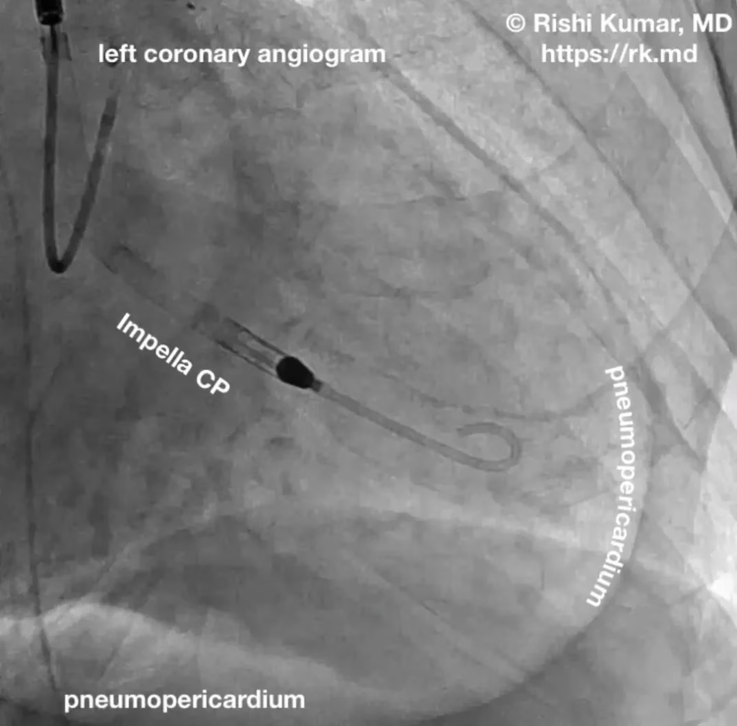
  • Pneumopericardium

    Pneumopericardium

    The heart is encased within a double-layered protective sac called the pericardium. Pneumopericardium occurs when air accumulates within this sac resulting in increased pressure around the heart. So how does air get there? Blunt or penetrating trauma to the chest can cause mediastinal disruption potentially leading to pneumopericardium. In addition,…

    Read More

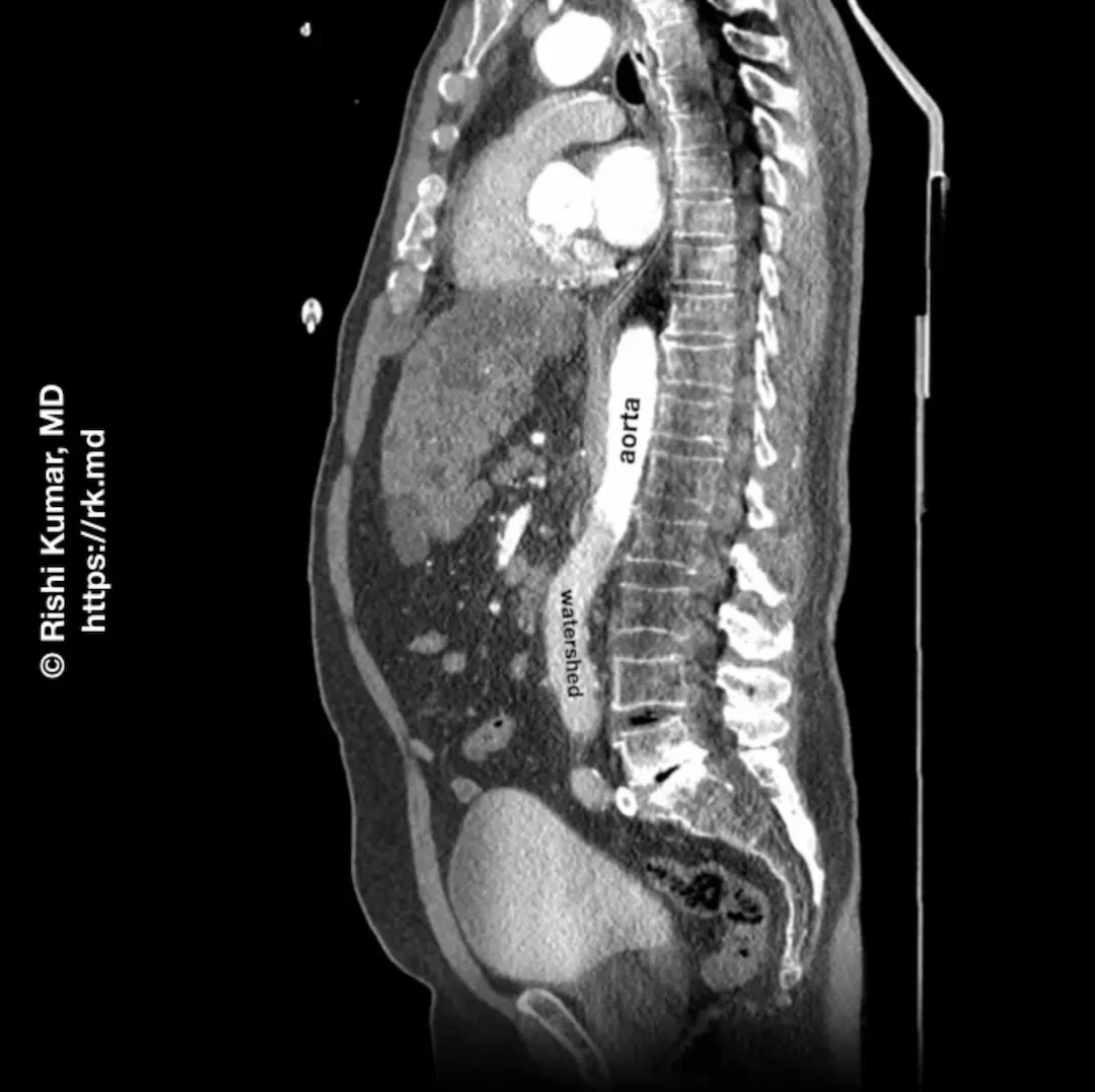
  • Peripheral VA-ECMO Watershed Area

    Peripheral VA-ECMO Watershed Area

    Peripheral venous-arterial (VA) ECMO, typically via the common femoral vein and artery, can be utilized as a bridge to recovery or decision in critically ill patients; however, this cannulation strategy can result in unique physiologic derangements like a watershed phenomenon. In this situation, perfusion occurs via antegrade cardiac output (blood…

    Read More

  • Calculating Cardiac Output With Volume-Time Integral (VTI)

    Calculating Cardiac Output With Volume-Time Integral (VTI)

    Cardiac output (CO) is mathematically calculated as the product of heart rate and stroke volume. It’s related to preload, afterload, and contractility. One echocardiographic technique I routinely use to estimate a patient’s stroke volume is the volume-time integral (VTI). The VTI method is based on the Doppler effect which relates…

    Read More

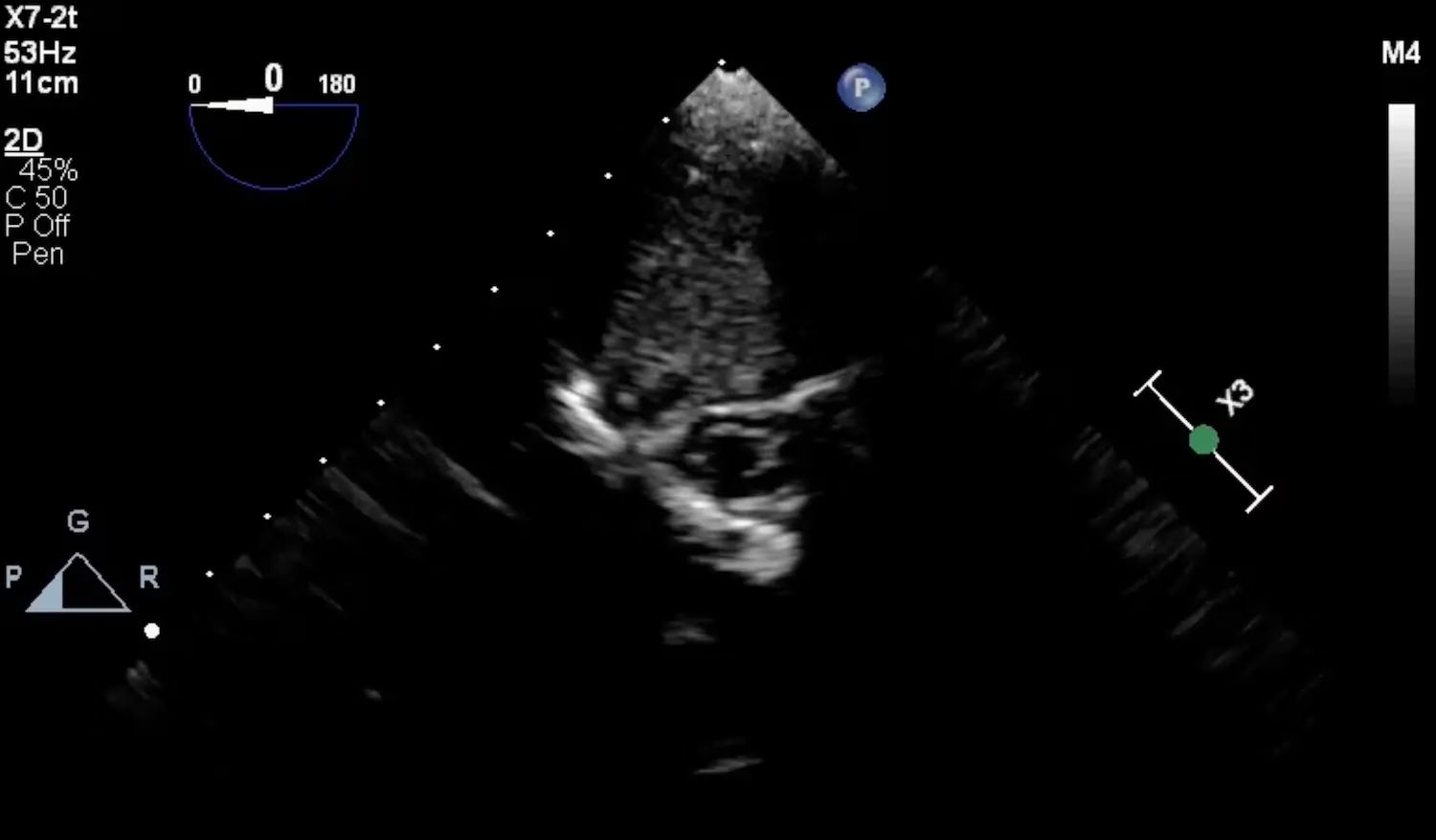
  • Spinal Cord On TEE

    Spinal Cord On TEE

    The heart is anterior to the esophagus, so the transesophageal echo (TEE) probe is facing anteriorly for the majority of the time; however, every once in a while when rotating clockwise/counter-clockwise, I’ll face posterior enough to visualize the spinal column. From a midesophageal region, one can advance and withdraw the…

    Read More

  • Superior Vena Cava (SVC) Collapsibility Index (CI)

    Superior Vena Cava (SVC) Collapsibility Index (CI)

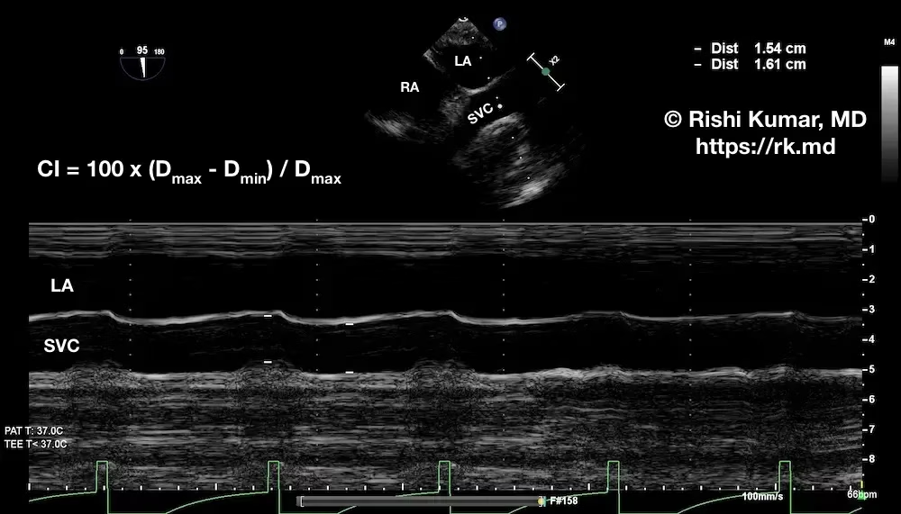
    To determine the superior vena cava (SVC) collapsibility index (CI), I use transesophageal echocardiography (TEE) to obtain a midesophageal bicaval view of the SVC in the long axis. I’ll then apply M-mode through the SVC to determine the maximum and minimum diameters (Dmax and Dmin, respectively) over the respiratory cycle.…

    Read More