
Fundoscopy examines the interior, back surface of the eye, including the retina, blood vessels, macula, and optic disc. Before learning pathology, one must first understand the “normal” anatomy of the retina. Let’s use a digital image of my right eye’s retinal structures for this purpose!
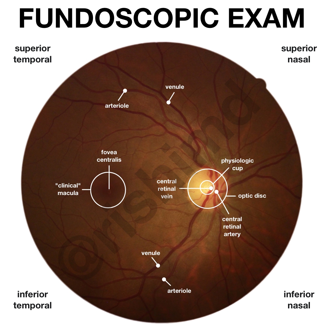
The visualized field can be divided into four quadrants – superior temporal (ST), superior nasal (SN), inferior temporal (IT), and inferior nasal (IN). In general, vessels emerge from the nasal side of the optic disc (veins are more prominent than arteries) and travel into all four quadrants as vascular arcades.
The retina is comprised of two primary photoreceptor cells – cones (concentrated in the macula, responsible for color vision) and rods (concentrated in the periphery, more sensitive than cones, responsible for night vision). The axons of retinal ganglion cells assemble to form the optic nerve (cranial nerve II) as it exits the optic disc. Because of this interruption in the retina, there are no photoreceptors at this point, leading to a physiologic blind spot. The optic cup-to-disc ratio should be < 0.5. The macula is located temporal to the optic disc and permits high-acuity, central, color (cone cells) vision.
Drop me a comment below with your questions!


